Written By Dr. Dawson November 2026
Some version of this visit happens in foot clinics every week. A patient with diabetes comes in because one shoe stopped fitting. The foot is swollen, warm, and a little red. It doesn't hurt, or it hurts far less than it should. Twice already, someone has looked at it: a sprain the first time, gout the second. There was no injury worth remembering. The other foot looks completely normal.
Nothing about that story sounds like an emergency. That is the problem.
That foot is often a Charcot foot, with bones and joints breaking down under intact skin in a foot that can't feel the damage. By the time it reaches a foot specialist, it has usually collected other diagnoses for weeks. Every one of those weeks, the patient walked on it.
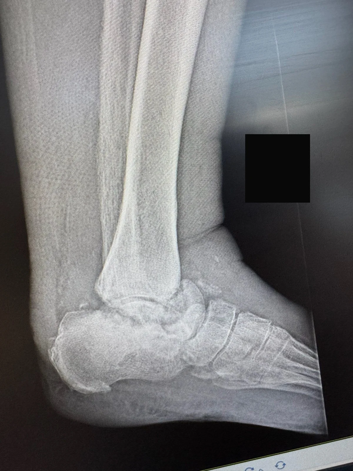
An ankle that has collapsed under Charcot. Nothing here sits where the blueprint put it, and every step now loads the wrong places.
TL;DR: Charcot neuroarthropathy is what happens when a foot loses its alarm system. Neuropathy silences the pain that normally forces rest, inflammation runs unchecked after a minor injury, and the bones and joints soften, fracture, and shift. The treatment that changes the outcome is unglamorous: immobilize the foot in a non-removable knee-high device, starting on suspicion, before imaging is finished.¹³ The honest clock is months, not weeks: a national audit found a median of 9 months to resolution with non-removable offloading.⁷ A hot, swollen foot in a person with diabetes and neuropathy is a get-seen-this-week problem. If there's an open sore, drainage, fever, or red streaking, it's a get-seen-today problem.
What is a Charcot foot?
Charcot neuroarthropathy, called Charcot arthropathy or Charcot neuro-osteoarthropathy, is the progressive breakdown of bones and joints in a foot with severe peripheral neuropathy.¹ Diabetes is now the most common cause of the nerve damage behind it.¹
Think of the foot as a building. The bones and joints are the frame. The nerves are the alarm system and wiring, reporting stress, injury, and position to the brain. A healthy frame complains when it's overloaded. Weight shifts, a shoe comes off, and the foot rests.
In a Charcot foot, the alarm fails first. Then the frame fails quietly, because nobody upstairs heard anything.
The skin usually stays closed. There's no cut, ulcer, or obvious entry point. A foot can be actively falling apart and look like a minor sprain that's taking its time. That intact skin is the most dangerous feature of this condition.
Why it happens: the alarm fails first
Neuropathy is the setup. Charcot can't happen in a foot with normal sensation. Years of diabetes damage the peripheral nerves until protective sensation is gone: the ability to feel a fracture, a stress injury, or a joint giving way. The patient isn't ignoring pain. The signal never arrives.
A trigger starts it. The match is usually small. In the UK CDUK audit of 288 cases, 36% recalled trauma in the prior 6 months, and 12% had recent surgery on the foot.⁷ A twisted ankle, a long walk in new shoes, a minor procedure. In a foot that can feel, that buys a limp and rest. In a neuropathic foot, it buys nothing. The patient keeps walking at full weight.
Inflammation does the demolition. The current model, laid out by Jeffcoate and Game in Diabetes Care, is an injury response critically distorted by nerve damage, an imperfect storm.² A normal injury triggers a controlled response: blood flow rises, bone temporarily softens, repairs begin, and pain enforces rest. In the neuropathic foot, neural controls on inflammation and blood flow are broken, pain enforces nothing, and the response overshoots. Pro-inflammatory cytokines, chemical messengers that tell immune and bone cells to act, drive a pathway called RANKL. That signal switches on osteoclasts, the cells that dissolve bone.³ Laboratory work confirms that this pathway is active and abnormal in Charcot tissue.⁴ Bone softens exactly when the patient loads it hardest.
Fractures accumulate, joints subluxate (slide partly out of place) or dislocate, and the architecture changes over weeks. The arch can collapse until the middle of the foot becomes the lowest point, a rocker-bottom shape.
Non-diabetic causes. Diabetes is the usual cause, but it isn't the only one. Any condition that takes away protective sensation can set the same table. Alcohol-related neuropathy is the non-diabetic cause I ask about most. Spinal cord conditions, leprosy, and other nerve diseases can do it too. The label matters less than whether the foot can still protect itself.¹
Who gets it. The best anchor is a prospective diabetes disease management cohort: 8.5 cases per 1,000 people with diabetes per year.⁵ A Danish registry letter reached the same broad conclusion: even in diabetes, this is uncommon.⁶ Without the neuropathy, Charcot can't happen. With it, a small trigger and time are enough.
🔗 Related Reading: Foot and Ankle Neuropathy: Myths, Scams, And What Actually Works: A plain-language tour of what neuropathy does to a foot, which advertised cures hold up, and what actually helps.
The four stages, and the one that matters most
Charcot is staged with the Eichenholtz system, described in his 1966 monograph and still the standard language today.⁸ ⁹
Stage 0 was added in 1990 because clinicians kept meeting patients whose feet were clearly in trouble while their X-rays were clean.⁸ It's the window that matters. A foot caught then and immobilized can heal in its original shape. A foot caught at stage 1 has usually started to deform.
Doctors also describe location with the Sanders and Frykberg patterns: pattern I in the forefoot, pattern II at the tarsometatarsal joints (the Lisfranc complex in the midfoot), pattern III in the midfoot joints just behind those, pattern IV at the ankle and subtalar joints, and pattern V in the heel bone.¹⁰ The midfoot patterns are the classic collapse sites. Location drives the treatment plan and surgical options.
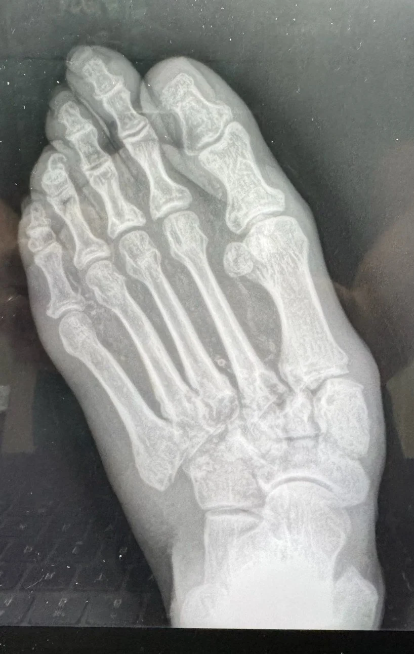
The midfoot is where Charcot does its most common damage. These joints used to hold up an arch.
🔗 Related Reading: Foot & Ankle Anatomy Explained: A Patient’s Guide to Bones, Joints, Nerves, Arteries: A quick map of the bones, joints, and terms used in this article, in plain language.
The diagnostic trap
Here's the statistic for every urgent care wall: in a 2022 systematic review, 53% of cases were delayed or misdiagnosed at first, and the average delay was nearly 3 months.¹¹
The disguises are consistent. The largest misdiagnosis category was skin and soft tissue infection, at 34.1% of delayed cases, followed by gout and arthritis, fracture and sprain, and deep vein thrombosis.¹¹
The delay isn't a paperwork problem. It's a shape problem. In a Swiss series of 71 cases managed with MRI, 13 cases had normal radiographs that were never cross-checked with MRI, and referral was postponed until 5 months after symptoms began. By then, fractures in the cortical bone, the hard outer shell of the bone, had occurred in 12 of them.¹² The X-ray said nothing was wrong. It was answering a later question. Meanwhile the foot kept failing under full daily load, and a foot that could have healed straight healed crooked.
The rule: In a patient with diabetes and neuropathy, a warm, swollen foot is Charcot until proven otherwise. Applied early, that sentence is worth more than any drug in this article.
How it is diagnosed
There's no single test that stamps this diagnosis. It's a clinical call, built from the exam, supported by imaging, and made early on purpose. The International Working Group on the Diabetic Foot (IWGDF) 2023 guideline is explicit: immobilize on clinical suspicion, before the imaging workup is complete.¹³ Waiting for certainty is how feet deform.
The exam. We compare both feet. The Charcot foot is warmer, usually measurably warmer. We use an infrared thermometer on matching spots, and a difference of 2 °C or more is the activity signal most clinics work with. That's a practice threshold, not a proven cutoff, and the guideline grades the temperature evidence as low certainty.¹³ ¹⁴ We look for swelling that stops mid-foot, redness without a wound, a lower arch, or a midfoot that feels unstable or boggy when stressed. We check for any break in the skin, because a break changes the problem. And we test sensation, because neuropathy is the entry requirement.
Imaging. Weight-bearing X-rays come first. At stage 1 and beyond they show fragmentation, debris, and joints out of place. At stage 0 they show nothing, and nothing is the finding that misleads people. When the X-ray is normal and the foot is hot, the guideline recommends MRI.¹³ In a study of 26 feet, MRI showed bone marrow edema, occult fractures, and joint effusion while radiographs were still normal.¹⁵
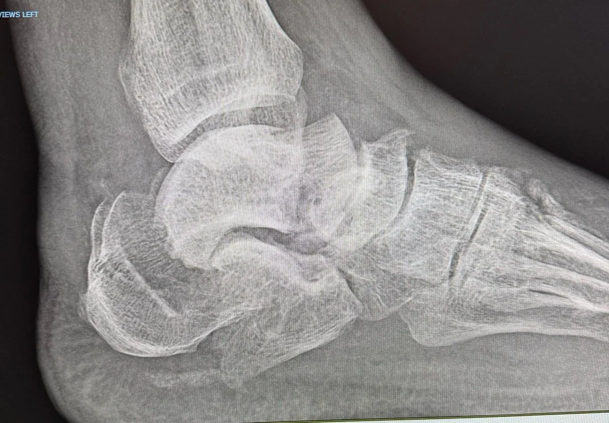
A heel bone that fractured without a fall. Load alone did this, applied day after day to bone that could not feel it failing.
Labs. Blood tests can't make this call. The IWGDF guideline recommends against using C-reactive protein, ESR (erythrocyte sedimentation rate), white cell count, and alkaline phosphatase to diagnose or exclude active Charcot when the skin is intact.¹³ Those markers rise in active Charcot with no infection anywhere, because the inflammation is real. It just isn't infectious.
Charcot or infection? Charcot inflammation happens under intact skin. Osteomyelitis, infection in the bone, almost always enters through an open ulcer. That fact does most of the sorting.
When there's an ulcer, a few findings move the needle. A large, deep ulcer makes bone infection much more likely, though it doesn't prove it. If a sterile probe passed gently through an ulcer touches bone, the probe-to-bone test is positive, and infection moves from possible to likely. A very high ESR raises concern, but it can't make the call alone because Charcot itself is inflammatory.¹⁶ ¹⁷
MRI catches most bone infection, and a normal MRI makes osteomyelitis much less likely. An abnormal MRI isn't a verdict, because Charcot and infection can both produce dramatic pictures. A probe that doesn't touch bone lowers concern but doesn't rule infection out.¹⁸
Sometimes both processes occupy the same foot. When the answer changes management, the definitive test is a bone biopsy, taken through clean skin away from any ulcer.¹⁹ Evidence grade: Moderate for the exam discriminators and MRI performance, thin almost everywhere else.¹⁴
The treatment ladder
Treatment is a ladder. The bottom rung does nearly all of the work.
Rung 1. Immobilize now, argue later. The IWGDF guideline names non-removable knee-high offloading as the gold-standard treatment for active Charcot, and the device goes on at the visit where suspicion lands, not when the MRI report returns.¹³ Removable walkers exist, and patients prefer them, but a removable device has a design flaw no engineering can fix: it comes off. The short walk to the kitchen counts.
Rung 2. The total contact cast. The workhorse is the total contact cast, a well-molded cast that spreads load across the foot and lower leg instead of concentrating it under the collapsing arch. It controls swelling, enforces rest the patient can't feel the need for, and holds the foot still while the hot phase burns out. Casts are changed serially, often weekly at first, because a swollen foot shrinks as it cools and a loose cast is a failed cast. Many patients bear weight in it with crutches or a walker. Some feet, particularly unstable ankle and hindfoot cases (the back part of the foot under and behind the ankle), need stricter unloading. Your surgeon sets that dial based on stability.
Now the honest clock. In the classic US specialty clinic cohort, 55 patients treated with serial total contact casting averaged 18.5 weeks in casts and reached prescription footwear at a mean of 28.3 weeks.²⁰ In the UK CDUK audit, median time to resolution was 9 months with non-removable offloading, versus 12 months without it.⁷ Plan on months. A foot that quiets in 6 weeks exists, but it's the exception that ruins everyone's expectations. Evidence grade for casting: Moderate.
What remission actually means. Remission isn't cured. It's quiet. The cast comes off when swelling and redness have resolved, the temperature difference stays under 2 °C across visits, and X-rays show consolidation. Then comes a graded transition through a temporary boot into definitive footwear, spread over weeks, with temperature checks continuing.
Rung 3. Footwear and bracing, for life. After remission, the foot keeps the shape it healed in, and that shape dictates protection. The IWGDF guideline recommends custom-made footwear and insoles that accommodate the foot's shape, and custom below-knee devices, such as a CROW (Charcot Restraint Orthotic Walker, essentially a rigid custom boot), when deformity or instability is present.¹³ This isn't a graduation. The shoe becomes part of the structure.
🔗 Related Reading: Corns and Calluses: What They Really Mean and How to Treat Them: How pressure and friction build callus over bony points, and how to manage skin before it breaks.
The drug detour, kept short
The logic was reasonable. Osteoclasts dissolve the bone, so block the osteoclasts. Bisphosphonates, calcitonin, and a shelf of related drugs all got their trial. Here's what the trials actually found.
The best of them, a double-blind randomized controlled trial of a single 90 mg pamidronate infusion in 39 patients, reduced bone turnover markers, symptoms, and foot temperature compared with placebo. It didn't show shorter immobilization or fewer deformities.²¹ Small randomized trials of oral alendronate and intranasal calcitonin moved surrogate markers only.²² ²³ Then the pattern broke the wrong way: in a pilot randomized trial of zoledronic acid, the treatment group needed longer immobilization, a median of 27 weeks versus 20 weeks.²⁴ The UK audit agreed: bisphosphonate use was associated with slower resolution, 12 months versus 10 months.⁷
The IWGDF guideline turned that record into a recommendation: don't use alendronate, pamidronate, zoledronate, calcitonin, parathyroid hormone, or methylprednisolone for active Charcot, and denosumab is suggested against as well.¹³
The drugs cooled the thermometer and quieted the lab report. The frame didn't care. A cooler foot that still collapses isn't a win. If someone offers an infusion as a shortcut around the cast, the trial record says the shortcut runs long. Evidence grade: Mixed for lab numbers, negative for every outcome a patient would actually choose.
Surgery: selected feet, real judgment
Surgery is the last rung of the ladder, and most Charcot feet never need it. When it does come up, the decision runs on four questions, and most of them get asked long before anyone opens an implant catalog.
How I sort the surgical foot. First, can the foot be kept plantigrade, stable, and closed in a cast or brace? Plantigrade means the sole can sit flat on the floor, with the heel and forefoot sharing load in a way a shoe or brace can work with. If the foot can stay plantigrade, surgery usually waits. Second, what's the skin doing? A closed, supple envelope gives us options; a tight, threatened, or ulcerated envelope narrows them. Third, is blood flow good enough to heal an incision and a fusion? Pulses, skin quality, and the overall vascular picture matter before reconstruction is promised. Fourth, is infection present? If it is, the sequence may need to change. Stability, skin, blood flow, and infection decide more cases than any implant catalog, and that sorting framework is the one the IWGDF guideline builds its surgical recommendations on.¹³
Shaving the bump. When a foot is stable and plantigrade but a bony prominence on the bottom keeps breaking the skin open, an exostectomy removes the prominence. It's the smallest operation in this article, and it treats a pressure problem, not the Charcot process itself. The systematic reviews support it as part of the standard algorithm.²⁶ ²⁷
Rebuilding the frame. When the foot is unstable or non-plantigrade, meaning it can't sit flat and bear weight in a brace, reconstruction fuses the collapsed joints into a straight, plantigrade shape. Modern reconstruction follows the superconstruct principles described by Sammarco, translated out of surgical shorthand: fuse past the damage, into joints that aren't involved, because hardware anchored in diseased bone fails; remove bone on purpose, shortening the foot so correction doesn't stretch the soft tissues to tearing; use the strongest fixation the soft tissue envelope can tolerate; and place it where the mechanical advantage is greatest.²⁵ Plates and screws inside the foot are one route. An external fixator is another, used when the skin is fragile or infection makes internal hardware a bad bet.
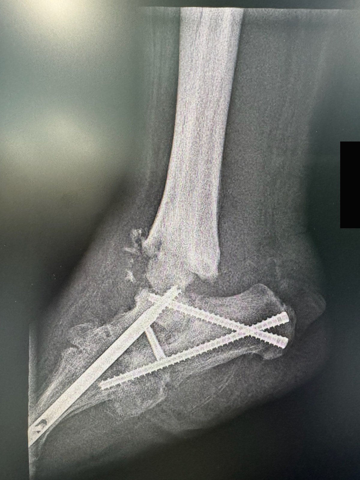
The screws held. The ankle above them dislocated anyway. Charcot does not respect a surgical plan, which is why follow-up after reconstruction is measured in years, not weeks.
Now the honesty paragraph. A systematic review examined 95 articles and found every included study was Level IV or V evidence: case series, no control groups, no randomization.²⁶ That doesn't mean surgery doesn't work. It means the literature can't prove how well it works, and outcomes ride heavily on selection and surgeon experience. In my clinic, surgery is the answer for a foot that can't be kept plantigrade, braced, and closed. It isn't the answer to impatience with a cast.
Operating while the foot is still hot. Some feet can be reconstructed while they're still hot. In the active phase the bones and joints are easier to move back into place, and for an unstable foot that's coming apart, waiting can mean rebuilding a worse deformity later. That isn't every foot. Most hot feet belong in a cast. Reconstruction during the active phase is a case-by-case call, made on stability, skin, blood flow, and the patient in front of me. The published support for operating early is thin and old, and it deserves to be sized honestly. In the best-known series, 14 patients with stage 1 midfoot Charcot chose early arthrodesis over casting, and all 14 achieved stability, union, and anatomic reduction, with no complications, back in regular shoes at a mean of 27 weeks, at a mean follow-up of 41 months.³⁶ One small series from 2000 shows the operation can work in a selected hot foot. It does not make it the default, and operating earlier does not make the decision casual: inflamed bone is soft, the tissues are swollen, and protected healing follows.
Amputation, said plainly. Everything above exists to prevent this. In a large VA cohort, overall amputation rates for Charcot and foot ulcer were similar, and Charcot without ulceration carried low risk. Ulceration transforms it. In patients under 65, Charcot plus ulcer carried 12 times the amputation risk of Charcot alone.³⁰ The open wound on the deformed foot is the enemy: the prominence ulcerates, the ulcer infects the bone, and the conversation changes. The counterweight is real too: with consistent offloading and footwear, the Swiss cohort's major amputation-free survival was 92.9% at a median follow-up of 5.2 years.²⁹ Amputation enters the discussion for uncontrolled bone infection, tissue loss, or a foot that can't be made plantigrade and kept closed. It's a salvage decision, not where this starts. For most patients who offload early and keep the skin closed, it isn't where this ends.
🔗 Related Reading: Your Complete Guide to Diabetic Foot Care: Protecting Your Feet for Life: The daily foot check, footwear rules, and warning signs that protect a diabetic foot for the long term.
Living with a Charcot foot
Recurrence. In a Swiss study of 57 feet followed for a mean of 47 months, Charcot reactivated in 23% of feet, at a mean of 27 months after immobilization ended. Feet that recurred had been immobilized for a shorter initial period, and the dominant predictor was non-compliance with offloading.²⁸ The strongest risk factor for doing this all again is not finishing the first time.
The other foot. It's now load-bearing for two. By the end of follow-up in the Swiss cohort, 32.1% of patients had Charcot involvement in both feet.²⁹ Your other foot has the same neuropathy, and while one foot is casted it takes double traffic. Protect it like it's already diagnosed: prescribed footwear on both feet, never barefoot, and the same daily inspection.
Daily habits, the short list. Look at both feet every day, tops and soles, with a mirror or a phone flashlight. Run a hand inside your shoes before they go on. Compare the warmth of both feet with the back of your hand, and if your clinic gave you a thermometer routine, keep it. An ulcer developed in 72.3% of the Swiss cohort over long-term follow-up, which is why inspection isn't a suggestion.²⁹ If you smoke, stop: smoking was associated with ulcer development and amputation in that cohort.²⁹
Call the same day if:
A new open sore appears on either foot, even a small one.
A foot that had cooled down gets warm, red, or swollen again.
There's drainage, odor, or a wet spot on the sock.
Red streaking spreads up the foot or leg.
You develop a fever of 100.4 °F (38 °C) or higher.
The foot's shape changes, or a cast feels loose, tight, or cracked.
The bigger picture, without flinching. A Charcot diagnosis travels with serious company. Population data link it with higher mortality than diabetes alone and with reduced life expectancy after an acute episode.³¹ ³² Those numbers aren't a personal countdown, and they aren't caused by the foot itself. The same vascular and metabolic disease is active everywhere, so the diagnosis should tighten the whole picture: heart risk, kidney checks, blood sugar, smoking. The foot is the loudest symptom. It isn't the only one.
Questions patients actually ask
How long will I be in a cast?
Months. The US specialty clinic cohort averaged 18.5 weeks in casts and about 28 weeks to prescription footwear.²⁰ Anyone who promises you 6 weeks is selling something.
Can I walk on it?
Many patients bear weight in a total contact cast, usually with crutches or a walker, and the clinic checks the cast as swelling falls. Unstable ankle and hindfoot cases get stricter rules. What you can't do is walk on it unprotected, including short trips that supposedly don't count. They're most of the load the foot sees.
Can it come back?
Yes. Recurrence was 23% at a mean of 27 months in one series, and the strongest predictor was stopping offloading early.²⁸ Finishing the full course, then staying in prescribed footwear, is the protection you control.
Can it happen to the other foot?
Yes. Bilateral involvement reached 32.1% by the end of long-term follow-up in the Swiss cohort.²⁹ Inspect and protect both feet from day one.
Will I lose my foot?
Charcot alone carries a low amputation risk. Charcot plus an open ulcer is the dangerous combination, with 12 times the risk of Charcot alone in patients under 65 in the VA data.³⁰ Keeping the skin closed isn't a detail. It's the whole game.
Can I prevent it?
No drug or procedure is proven to prevent Charcot. The levers are unglamorous: prescribed footwear once neuropathy is established, daily inspection, temperature checks if your clinic recommends them, and treating every hot, swollen, painless foot as a this-week emergency. Early is the only shortcut this condition offers.
The Bottom Line
A Charcot foot is a building whose alarm system will never be rewired. The nerves aren't coming back, and no infusion changes that. So the owner becomes the alarm: a daily walk-through, hand on the walls, checking for heat, looking for the crack in the plaster before it becomes the hole in the floor. Pair that habit with a cast applied in the first weeks instead of the fourth month, and the building stands, plantigrade, shoeable, and closed. That's the strategy. It has no marketing department, and it works.
Recommended products for the daily routine
These items are listed for education and reference only. They are not a substitute for medical advice. Please see your podiatrist for specific recommendations. I may receive a commission if you purchase through these links, at no additional cost to you.
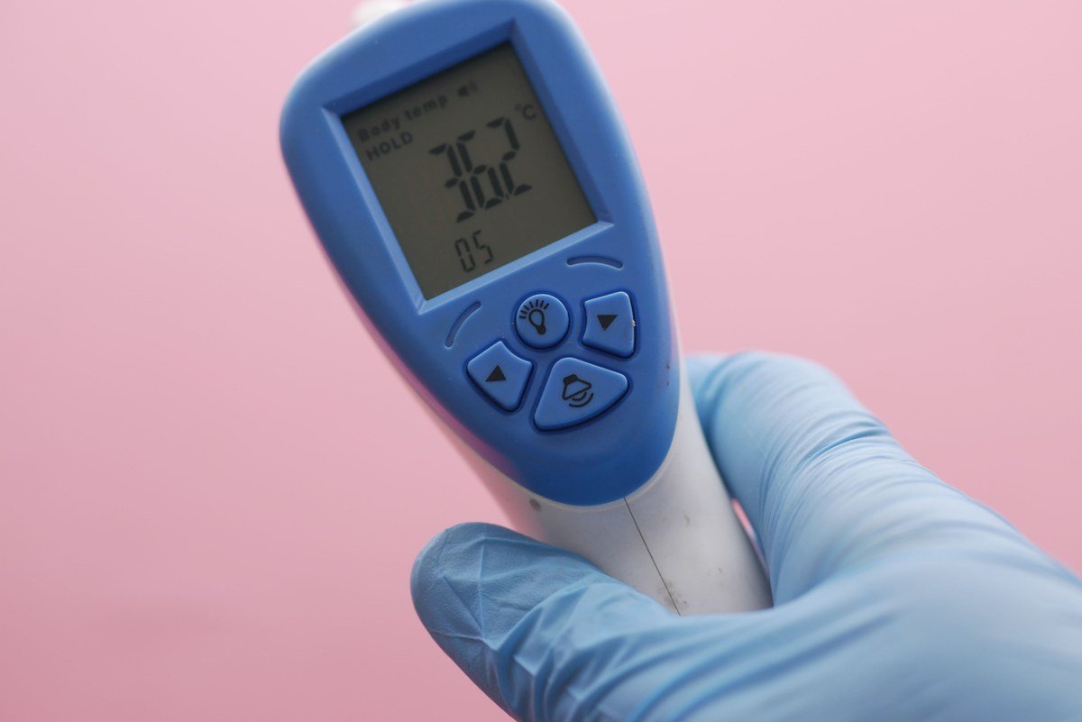
The whole test is a comparison: same spot, other foot, write it down.
ThermoWorks WAND Clinical Non-Contact Thermometer. This is the one device here with randomized trial support. In high-risk patients with diabetes, home skin temperature monitoring reduced foot complications and ulcer recurrence when a hot spot triggered the important part: less activity and a call to the care team.³³ ³⁴ ³⁵ The trials used a handheld infrared device called the TempTouch, built for this job and no longer sold. The work itself is simple: read the temperature at matching spots on both feet and compare. The WAND Clinical is an FDA-cleared clinical thermometer, and it publishes accuracy at the temperatures feet actually run, which are cooler than a forehead. That is why it is the pick here. Use it on the same spots every time and write the numbers down. In the trials, a difference greater than 2.2 °C at the same spot was the action threshold. Your clinic's active-treatment threshold wins if it differs. A thermometer doesn't treat Charcot, and it never substitutes for a cast.
Sammons Preston Telescoping Self-Examination Mirror. You can't protect a sole you never see. This mirror's handle extends from 6 to 18 inches, so the sole comes to you instead of the other way around, and one side magnifies 2x for a closer look. Use it daily, in good light, sitting comfortably: check the bottom of both feet for redness, blisters, cracks, and new sores, and ask for help when the angle won't cooperate.
Dr. Comfort Diabetic Crew Socks. Choose socks with no raised seam across the toes and a top that doesn't bite into the calf. These are built that way, with seamless construction and a non-binding top. Change them daily, and check for a wet spot when you take them off, because drainage can show on fabric before you notice it on skin. Socks are protection, not treatment. They reduce rubbing; they don't offload a hot foot, and they don't make barefoot walking safe once they come off.
References
1. Rogers LC, Frykberg RG, Armstrong DG, Boulton AJM, Edmonds M, Van GH, Hartemann A, Game F, Jeffcoate W, Jirkovska A, Jude E, Morbach S, Morrison WB, Pinzur M, Pitocco D, Sanders L, Wukich DK, Uccioli L. The Charcot foot in diabetes. Diabetes Care. 2011;34(9):2123-2129. DOI: 10.2337/dc11-0844. PMID: 21868781.
2. Jeffcoate W, Game F. The Charcot foot reflects a response to injury that is critically distorted by preexisting nerve damage: an imperfect storm. Diabetes Care. 2022;45(7):1691-1697. DOI: 10.2337/dc21-2508. PMID: 35796768.
3. Jeffcoate WJ, Game FL, Cavanagh PR. The role of proinflammatory cytokines in the cause of neuropathic osteoarthropathy (acute Charcot foot) in diabetes. The Lancet. 2005;366(9502):2058-2061. DOI: 10.1016/S0140-6736(05)67029-8. PMID: 16338454.
4. Ndip A, Williams A, Jude EB, Serracino-Inglott F, Richardson S, Smyth JV, Boulton AJM, Alexander MY. The RANKL/RANK/OPG signaling pathway mediates medial arterial calcification in diabetic Charcot neuroarthropathy. Diabetes. 2011;60(8):2187-2196. DOI: 10.2337/db10-1220. PMID: 21659498.
5. Lavery LA, Armstrong DG, Wunderlich RP, Tredwell J, Boulton AJM. Diabetic foot syndrome: evaluating the prevalence and incidence of foot pathology in Mexican Americans and non-Hispanic whites from a diabetes disease management cohort. Diabetes Care. 2003;26(5):1435-1438. DOI: 10.2337/diacare.26.5.1435. PMID: 12716801.
6. Svendsen OL, Rabe OC, Winter-Jensen M, Højgaard-Allin K. How common is the rare Charcot foot in patients with diabetes? Diabetes Care. 2021;44(4):e62-e63. DOI: 10.2337/dc20-2590. PMID: 33526427.
7. Game FL, Catlow R, Jones GR, Edmonds ME, Jude EB, Rayman G, Jeffcoate WJ. Audit of acute Charcot's disease in the UK: the CDUK study. Diabetologia. 2012;55(1):32-35. DOI: 10.1007/s00125-011-2354-7. PMID: 22065087.
8. Rosenbaum AJ, DiPreta JA. Classifications in brief: Eichenholtz classification of Charcot arthropathy. Clinical Orthopaedics and Related Research. 2015;473(3):1168-1171. DOI: 10.1007/s11999-014-4059-y. PMID: 25413713.
9. Eichenholtz SN. Charcot Joints. Springfield, IL: Charles C. Thomas; 1966.
10. Sanders LJ, Frykberg RG. Diabetic neuropathic osteoarthropathy: the Charcot foot. In: Frykberg RG, ed. The High Risk Foot in Diabetes Mellitus. New York: Churchill Livingstone; 1991:297-338.
11. Korst GS, Ratliff HT, Torian J, Jimoh RO, Jupiter DC. Delayed diagnosis of Charcot foot: a systematic review. The Journal of Foot and Ankle Surgery. 2022;61(5):1109-1113. DOI: 10.1053/j.jfas.2022.01.008. PMID: 35236617.
12. Chantelau EA, Richter A. The acute diabetic Charcot foot managed on the basis of magnetic resonance imaging: a review of 71 cases. Swiss Medical Weekly. 2013;143:w13831. DOI: 10.4414/smw.2013.13831. PMID: 23897135.
13. Wukich DK, Schaper NC, Gooday C, Bal A, Bem R, Chhabra A, Hastings M, Holmes C, Petrova NL, Santini Araujo MG, Senneville E, Raspovic KM. Guidelines on the diagnosis and treatment of active Charcot neuro-osteoarthropathy in persons with diabetes mellitus (IWGDF 2023). Diabetes/Metabolism Research and Reviews. 2024;40(3):e3646. DOI: 10.1002/dmrr.3646. PMID: 37218537.
14. Raspovic KM, Schaper NC, Gooday C, Bal A, Bem R, Chhabra A, Hastings M, Holmes C, Petrova NL, Santini Araujo MG, Senneville E, Wukich DK. Diagnosis and treatment of active Charcot neuro-osteoarthropathy in persons with diabetes mellitus: a systematic review. Diabetes/Metabolism Research and Reviews. 2024;40(3):e3653. DOI: 10.1002/dmrr.3653. PMID: 37179484.
15. Chantelau E, Poll LW. Evaluation of the diabetic Charcot foot by MR imaging or plain radiography: an observational study. Experimental and Clinical Endocrinology & Diabetes. 2006;114(8):428-431. DOI: 10.1055/s-2006-924229. PMID: 17039424.
16. Butalia S, Palda VA, Sargeant RJ, Detsky AS, Mourad O. Does this patient with diabetes have osteomyelitis of the lower extremity? JAMA. 2008;299(7):806-813. DOI: 10.1001/jama.299.7.806. PMID: 18285592.
17. Grayson ML, Gibbons GW, Balogh K, Levin E, Karchmer AW. Probing to bone in infected pedal ulcers: a clinical sign of underlying osteomyelitis in diabetic patients. JAMA. 1995;273(9):721-723. DOI: 10.1001/jama.1995.03520330051036. PMID: 7853630.
18. Dinh MT, Abad CL, Safdar N. Diagnostic accuracy of the physical examination and imaging tests for osteomyelitis underlying diabetic foot ulcers: meta-analysis. Clinical Infectious Diseases. 2008;47(4):519-527. DOI: 10.1086/590011. PMID: 18611152.
19. Donegan R, Sumpio B, Blume PA. Charcot foot and ankle with osteomyelitis. Diabetic Foot & Ankle. 2013;4:21361. DOI: 10.3402/dfa.v4i0.21361. PMID: 24098835.
20. Armstrong DG, Todd WF, Lavery LA, Harkless LB, Bushman TR. The natural history of acute Charcot's arthropathy in a diabetic foot specialty clinic. Journal of the American Podiatric Medical Association. 1997;87(6):272-278. DOI: 10.7547/87507315-87-6-272. PMID: 9198348.
21. Jude EB, Selby PL, Burgess J, Lilleystone P, Mawer EB, Page SR, Donohoe M, Foster AV, Edmonds ME, Boulton AJM. Bisphosphonates in the treatment of Charcot neuroarthropathy: a double-blind randomised controlled trial. Diabetologia. 2001;44:2032-2037. DOI: 10.1007/s001250100008. PMID: 11719835.
22. Pitocco D, Ruotolo V, Caputo S, Mancini L, Collina CM, Manto A, Caradonna P, Ghirlanda G. Six-month treatment with alendronate in acute Charcot neuroarthropathy: a randomized controlled trial. Diabetes Care. 2005;28(5):1214-1215. DOI: 10.2337/diacare.28.5.1214. PMID: 15855594.
23. Bem R, Jirkovská A, Fejfarová V, Skibová J, Jude EB. Intranasal calcitonin in the treatment of acute Charcot neuroosteoarthropathy: a randomized controlled trial. Diabetes Care. 2006;29(6):1392-1394. DOI: 10.2337/dc06-0376. PMID: 16732029.
24. Pakarinen TK, Laine HJ, Mäenpää H, Mattila P, Lahtela J. The effect of zoledronic acid on the clinical resolution of Charcot neuroarthropathy: a pilot randomized controlled trial. Diabetes Care. 2011;34(7):1514-1516. DOI: 10.2337/dc11-0396. PMID: 21593295.
25. Sammarco VJ. Superconstructs in the treatment of Charcot foot deformity: plantar plating, locked plating, and axial screw fixation. Foot and Ankle Clinics of North America. 2009;14(3):393-407. DOI: 10.1016/j.fcl.2009.04.004. PMID: 19712882.
26. Lowery NJ, Woods JB, Armstrong DG, Wukich DK. Surgical management of Charcot neuroarthropathy of the foot and ankle: a systematic review. Foot & Ankle International. 2012;33(2):113-121. DOI: 10.3113/FAI.2012.0113. PMID: 22381342.
27. Schneekloth BJ, Lowery NJ, Wukich DK. Charcot neuroarthropathy in patients with diabetes: an updated systematic review of surgical management. The Journal of Foot and Ankle Surgery. 2016;55(3):586-590. DOI: 10.1053/j.jfas.2015.12.001. PMID: 26810129.
28. Osterhoff G, Böni T, Berli M. Recurrence of acute Charcot neuropathic osteoarthropathy after conservative treatment. Foot & Ankle International. 2013;34(3):359-364. DOI: 10.1177/1071100712464957. PMID: 23520293.
29. Gratwohl V, Jentzsch T, Schöni M, Kaiser D, Berli MC, Böni T, Waibel FWA. Long-term follow-up of conservative treatment of Charcot feet. Archives of Orthopaedic and Trauma Surgery. 2022;142(10):2553-2566. DOI: 10.1007/s00402-021-03881-5. PMID: 33829302.
30. Sohn MW, Stuck RM, Pinzur M, Lee TA, Budiman-Mak E. Lower-extremity amputation risk after Charcot arthropathy and diabetic foot ulcer. Diabetes Care. 2010;33(1):98-100. DOI: 10.2337/dc09-1497. PMID: 19825822.
31. Sohn MW, Lee TA, Stuck RM, Frykberg RG, Budiman-Mak E. Mortality risk of Charcot arthropathy compared with that of diabetic foot ulcer and diabetes alone. Diabetes Care. 2009;32(5):816-821. DOI: 10.2337/dc08-1695. PMID: 19196882.
32. van Baal J, Hubbard R, Game F, Jeffcoate W. Mortality associated with acute Charcot foot and neuropathic foot ulceration. Diabetes Care. 2010;33(5):1086-1089. DOI: 10.2337/dc09-1428. PMID: 20185744.
33. Lavery LA, Higgins KR, Lanctot DR, Constantinides GP, Zamorano RG, Armstrong DG, Athanasiou KA, Agrawal CM. Home monitoring of foot skin temperatures to prevent ulceration. Diabetes Care. 2004;27(11):2642-2647. DOI: 10.2337/diacare.27.11.2642. PMID: 15504999.
34. Lavery LA, Higgins KR, Lanctot DR, Constantinides GP, Zamorano RG, Athanasiou KA, Armstrong DG, Agrawal CM. Preventing diabetic foot ulcer recurrence in high-risk patients: use of temperature monitoring as a self-assessment tool. Diabetes Care. 2007;30(1):14-20. DOI: 10.2337/dc06-1600. PMID: 17192326.
35. Armstrong DG, Holtz-Neiderer K, Wendel C, Mohler MJ, Kimbriel HR, Lavery LA. Skin temperature monitoring reduces the risk for diabetic foot ulceration in high-risk patients. The American Journal of Medicine. 2007;120(12):1042-1046. DOI: 10.1016/j.amjmed.2007.06.028. PMID: 18060924.
36. Simon SR, Tejwani SG, Wilson DL, Santner TJ, Denniston NL. Arthrodesis as an early alternative to nonoperative management of Charcot arthropathy of the diabetic foot. Journal of Bone and Joint Surgery American Volume. 2000;82-A(7):939-950. DOI: 10.2106/00004623-200007000-00005. PMID: 10901308.
Dr. Dawson
Hi, I'm Dr. Dawson, DPM, FACFAS, DABFAS, DABPM, a double board-certified foot and ankle surgeon. I spend most of my week in clinic and the operating room, and a surprising amount of the rest explaining why the $8 foam insert at the drugstore is not an orthotic or why a viral trend didn't do what they hyped it to. I started Feet Made Simple™ because patients deserve foot and ankle information built on peer-reviewed research, not product hype. Every post is written by me, fully referenced, and honest about where the evidence is strong and where it isn't. Biomechanics nerd at heart. Based in Charlotte, NC.