
Plantar Fasciitis: A Podiatrist’s Guide to Diagnosis & Evidence-Based Management
Plantar fasciitis remains the most common cause of plantar heel pain in adults, afflicting roughly 10 % of the population during their lifetime and prompting more than two million office visits each year.¹ Pathology studies have consistently shown the issue is more a ‘degenerative fasciosis’ — meaning the plantar fascia is weakening and breaking down² not just swelling — highlighting the need for mechanical rather than purely anti-inflammatory interventions. Therefore, I want you to fix the underlying problem and imbalance, not just ibuprofen it away.

Anatomic Overview of Plantar Fascia
Showing inflammation and tearing near the calcaneus insertion point
Pathophysiology & Clinical Presentation
The plantar fascia, a dense fibrous band running from the medial calcaneal tuberosity to the proximal phalanges, endures repetitive loading through your thousands of steps each day. Micro-tears and disorganized collagen trigger the classic symptom trio:
First-step pain – sharp, stabbing discomfort with initial weight-bearing after rest or sleep.
Warm-up effect – pain eases or even goes away after several minutes of ambulation, then returns with prolonged activity or standing.
Compensatory gait changes – chronic cases may refer stress to the knees, hips, or lumbar spine. Most often the inner knee and IT bands.
Related heel and forefoot topics: Morning heel pain often overlaps with Achilles tendinopathy. If pain sits under the big toe joint, read my guide to sesamoiditis.
Predisposing Factors
Anatomic / biomechanical:
Pes planus, pes cavus, limited ankle dorsiflexion, gastrocnemius-soleus tightness
These alter load distribution, increasing strain across the fascia
Activity-related
Distance running, occupations with prolonged standing, recent increase in activity. Get a little too carried away dancing at that last wedding?
Leading to repetitive micro-trauma and even minor tears in the fascia
Systemic / demographic
Elevated BMI, age 40–60 years, rapid weight gain
Causing increased plantar load and reduced tissue resilience
Pediatric note: True plantar fasciitis is uncommon in children; rule out calcaneal apophysitis (Sever’s disease) or progressing/worsening pain secondary to flatfoot.
Conservative Management (≈ 95 % Success Rate)
1. Structured Stretching Program³
Gastrocnemius stretch: knee extended, 30-second hold, three reps, twice daily. Using a wedge device can help stretch in the house using a wall and body weight.
Plantar fascia stretch: dorsiflex hallux and toes for 10–15 seconds before first steps.
Eight weeks of adherence yields significant pain reduction.
2. Footwear Optimization & Orthoses⁴
Replace worn footwear; choose models with firm heel counters and midfoot stability.
Even around the house, avoid flatfoot walking when the heel is especially painful. Using a rocker bottom style shoe like the OOfos can help but any well padded, supportive house slipper can help too
Begin with over-the-counter contoured insoles; escalate to custom orthoses after 6–8 weeks if needed.
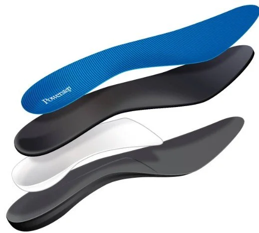
Anatomy of an OTC orthotic
Not ALL orthotics are created equal. Especially true for OTC orthotics. Make sure the device you buy has the supportive inner shell seen here in white.
(Pictured is the Powerstep brand, widely accessible online, highly regarded by many Podiatrists)
3. Night-Splint Therapy⁵
Dorsiflexion night splints come in two main varieties. Static and Adjustable. Static splints are used to sleep through the night and keep the Achilles tendon from getting tight while sleeping. They usually look like socks with a tether between the leg and the foot. Adjustable splints are more rigid and are used to get a deeper stretch that you use for 30-60 minutes a day.

Adjustable Night Splint
Used to gently stretch the Achilles tendon over a long period of time
4. Cryotherapy & Self-Massage
Roll a frozen water bottle beneath the arch for 2-5 minutes post-activity to provide short-term pain relief and gentle myofascial release.
5. Activity Modification
Substitute high-impact routines with cycling or swimming; take short walking or sitting breaks to offset prolonged standing.
More Invasive Options for Non-Healing Cases
Corticosteroid injections⁷
Usually short-term relief from pain; monitor for rare fascia rupture
Used for severe focal pain limiting rehab, does NOT cure plantar fasciitis
Minimally Invasive plantar fasciotomy⁸
Favorable outcomes in systematic reviews; however, < 5 % of patients go on to require surgery of any kind
Used for debilitating or non-improving pain after ≥ 6 months of evidence-based care
In my clinic this is done in the office setting and requires 1 stitch with the patient walking in regular shoes the same day!
Prognosis & Follow-Up
Most patients achieve meaningful improvement within 2–6 months; full resolution in >95% with conservative care alone. Clear communication of this timeline enhances adherence and satisfaction.
Referral to Podiatrist & Red-Flag Criteria
Persistent symptoms after 8 weeks of guideline-concordant therapy
Progressive neurologic signs (numbness, paresthesia). Could be a sign of a common mimic called Tarsal Tunnel Syndrome.
Atypical features suggestive of stress fracture or systemic inflammatory disease. Usually manifests opposite of plantar fasciitis where pain is better after rest and worse with activity.
Pediatric Note: Heel pain persisting > 2 weeks is a cause for concern. Kiddos usually bounce back from mild injuries quickly and don’t usually get wear and tear injuries as often as adults
Early Podiatrist evaluation expedites advanced imaging and alternative diagnoses when warranted.
Plantar Fasciitis FAQ
How long does plantar fasciitis take to heal?
Most people improve within twelve weeks with consistent loading, calf mobility, and shoe support. Morning symptoms often fade first, then end‑of‑day soreness follows.¹
Do I need an MRI or ultrasound?
Usually no. Diagnosis is clinical. Imaging is considered if pain does not improve after a focused program or if red flags suggest another diagnosis.¹
Are cortisone shots required?
Not usually. Injections may help short term but do not replace a progressive loading plan, footwear changes, and night positioning.¹
¹ References align with the main article’s citations from peer‑reviewed sources including JFAS, APMA, and AOFAS.
🦶 Recommended Products for Plantar Fasciitis
These items are listed for education and reference only. They are not a substitute for medical advice. Please see your podiatrist for specific recommendations. I may receive a commission if you purchase through these links, at no additional cost to you.
👟 Shoes
- Brooks Ghost Max 3 – Cushioned maximalist shoe with a rocker sole to offload heel pain
- Hoka Bondi 9 – Plush maximalist road shoe for shock absorption
- Topo Atmos – Maximalist shoe with wide forefoot for comfort and stability
🩴 Sandals
- OOfos Recovery Sandal – Rocker bottom relief sandal for post-activity comfort
🦾 Orthotics
- Powerstep Pinnacle Maxx – General purpose orthotic with firm heel control
🧰 Rehab Tools
- Rigid Night Splint – Keeps the plantar fascia stretched overnight
- Wedge Stretch Device – Simple calf and plantar fascia stretching tool
- Tuli Heel Cups – Shock-absorbing heel cups to offload pressure
- Theraband Resistance Bands – Bundle for ankle and foot strengthening
🧴 Skin & Nail Care
- Epsom Salts – Classic soak for sore or inflamed heels
References
Singh B, Yadav CP, Chakravarthy M, et al. Plantar fasciopathy: a current concepts review. EFORT Open Reviews. 2018;3(8):485-494. doi:10.1302/2058-5241.3.180018
Abdelkader NA, Elsayed S. Plantar fasciitis: an updated review. Life (Basel). 2024;14(2):173. doi:10.3390/life14020173
Pollack Y, Shashua A. The effectiveness of mechanical treatment for plantar fasciitis: a systematic review. Journal of Back and Musculoskeletal Rehabilitation. 2020;33(3):339-353. doi:10.3233/BMR-181442
Wapner KL, Sharkey PF. The use of night splints for treatment of recalcitrant plantar fasciitis. Foot & Ankle. 1991;12(3):135-137. doi:10.1177/107110079101200303
Gollwitzer H, Schmitz C, Guggenberger R, et al. Extracorporeal shock-wave therapy for chronic painful heel syndrome: a prospective, double-blind, randomized trial assessing the efficacy of a new electromagnetic shock-wave device. Journal of Foot & Ankle Surgery. 2007;46(5):348-357. doi:10.1053/j.jfas.2007.06.008
Li Z, Zhang X, Guo T, et al. Ultrasound- versus palpation-guided injection of corticosteroid for plantar fasciitis: a meta-analysis. PLoS ONE. 2014;9(3):e92671. doi:10.1371/journal.pone.0092671
Jerosch J. Endoscopic plantar fasciotomy versus open approach: a comparative study. Knee Surgery, Sports Traumatology, Arthroscopy. 2004;12(1):82-86. doi:10.1007/s00167-003-0442-4
Latt LD, Jaffar-Jezik L, Muz B, et al. Evaluation and treatment of chronic plantar fasciitis. Foot & Ankle Orthopaedics. 2020;5(1):2473011419896763. doi:10.1177/2473011419896763
Petraglia F, Papa S, Zaffagnini S, et al. Plantar fasciitis in athletes: diagnostic and treatment strategies. A systematic review. Muscles, Ligaments and Tendons Journal. 2017;7(1):107-118. doi:10.11138/mltj/2017.7.1.107
Riddle DL, Schappert SM. Volume of ambulatory care visits and patterns of care for patients diagnosed with plantar fasciitis: a national study of medical doctors. Foot & Ankle International. 2004;25(5):303-310. doi:10.1177/107110070402500505
Thomas JL, Christensen JC, Kravitz SR, et al. The diagnosis and treatment of heel pain: a clinical practice guideline—revision 2010. Journal of Foot & Ankle Surgery. 2010;49(3 Suppl):S1-S19. doi:10.1053/j.jfas.2010.01.001
Irving DB, Cook JL, Young MA, Menz HB. Obesity and pronated foot type may increase the risk of chronic plantar heel pain: a matched case-control study. BMC Musculoskeletal Disorders. 2007;8:41. doi:10.1186/1471-2474-8-41
Labovitz JM, Yu J, Kim C, DiMasi F. The relationship between gastrocnemius tightness and plantar fasciitis. Foot & Ankle Specialist. 2011;4(3):141-144. doi:10.1177/1938640011404670
Young CC, Rutherford DS, Niedfeldt MW. Treatment of plantar fasciitis. American Family Physician. 2005;72(11):2237-2242.