Written by Dr. Zack Dawson, DPM, foot and ankle surgeon · Last updated November 2026
It usually starts with a sound you feel more than hear. Your foot rolls on a curb, twists on the stairs, or plants wrong coming down from a ladder, and something gives. The pain is immediate. The ankle swells fast, sometimes within minutes, and putting weight on it ranges from miserable to impossible. In the bad ones the foot sits at an angle it should not sit at. By the time I meet most ankle fracture patients, they have already had the X-ray, the splint, and the same three questions. Is it broken? Do I need surgery? And when can I walk on it? This guide walks through all three, including the two fixation options I use most, the fibular nail and posterior plating, and what recovery actually looks like.
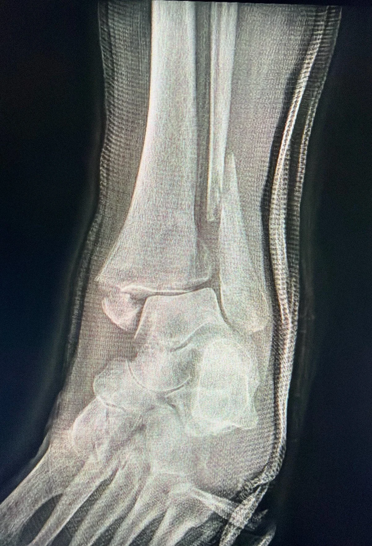
An injury film of a broken ankle in its ER splint. The outer bone is displaced, a piece of the inner ankle is broken, and the space between the leg bones has widened. That combination does not heal straight in a cast.
📌 TL;DR
A broken ankle means one or more of the three bones that form the ankle joint have cracked, and the pattern decides the treatment. Stable fractures with the joint still lined up can heal in a cast or boot, sometimes in as little as 3 weeks. Unstable fractures need surgery to put the joint back together, because a joint that heals crooked wears out. Two of the fixation choices inside that surgery are a plate on the outer bone or a rod down its center called a fibular nail, and for nails in otherwise stable patterns (secure fixation, no screw across the syndesmosis, a solid back fragment) I allow walking in a boot right away. A fragment at the back of the shin bone, the posterior malleolus, gets fixed through the back of the ankle when its shape calls for it, a decision made from the CT scan rather than the old size rule. Bone healing takes about 6 to 8 weeks. Getting your motion, strength, and normal life back takes months, and rehab is where that happens.
What Actually Broke
The ankle is a socket. The shin bone, the tibia, sits on top of the talus, the bone that transfers your weight into the foot, and the thin outer leg bone, the fibula, forms the outside wall. The bumps you can feel on either side are the ends of those bones, each one a malleolus. Inside is the medial malleolus, the end of the tibia. Outside is the lateral malleolus, the end of the fibula. The back edge of the tibia is the posterior malleolus.
Fracture patterns are named by how many of those walls broke.
Lateral malleolus fracture. Only the outer bone broke. This is the most common pattern, and the one that sometimes heals without surgery.
Isolated medial malleolus fracture. Only the inner bone broke, with no fracture on the outer side. This one is uncommon, and it earns a closer look, because it can travel with a break high on the fibula up near the knee, called a Maisonneuve fracture, and a torn syndesmosis, so the whole leg gets examined. When the ankle stays stable and the fragment is nondisplaced, it follows the same path as a stable outer bone fracture and heals without surgery.¹⁵
Bimalleolar fracture. The outer and inner bones are both broken.
Trimalleolar fracture. All three are broken, including the posterior malleolus at the back.
Fracture with syndesmosis injury. The ligaments binding the tibia and fibula together just above the joint, called the syndesmosis, are torn as well. The socket itself becomes loose, and that looseness matters as much as the bones.
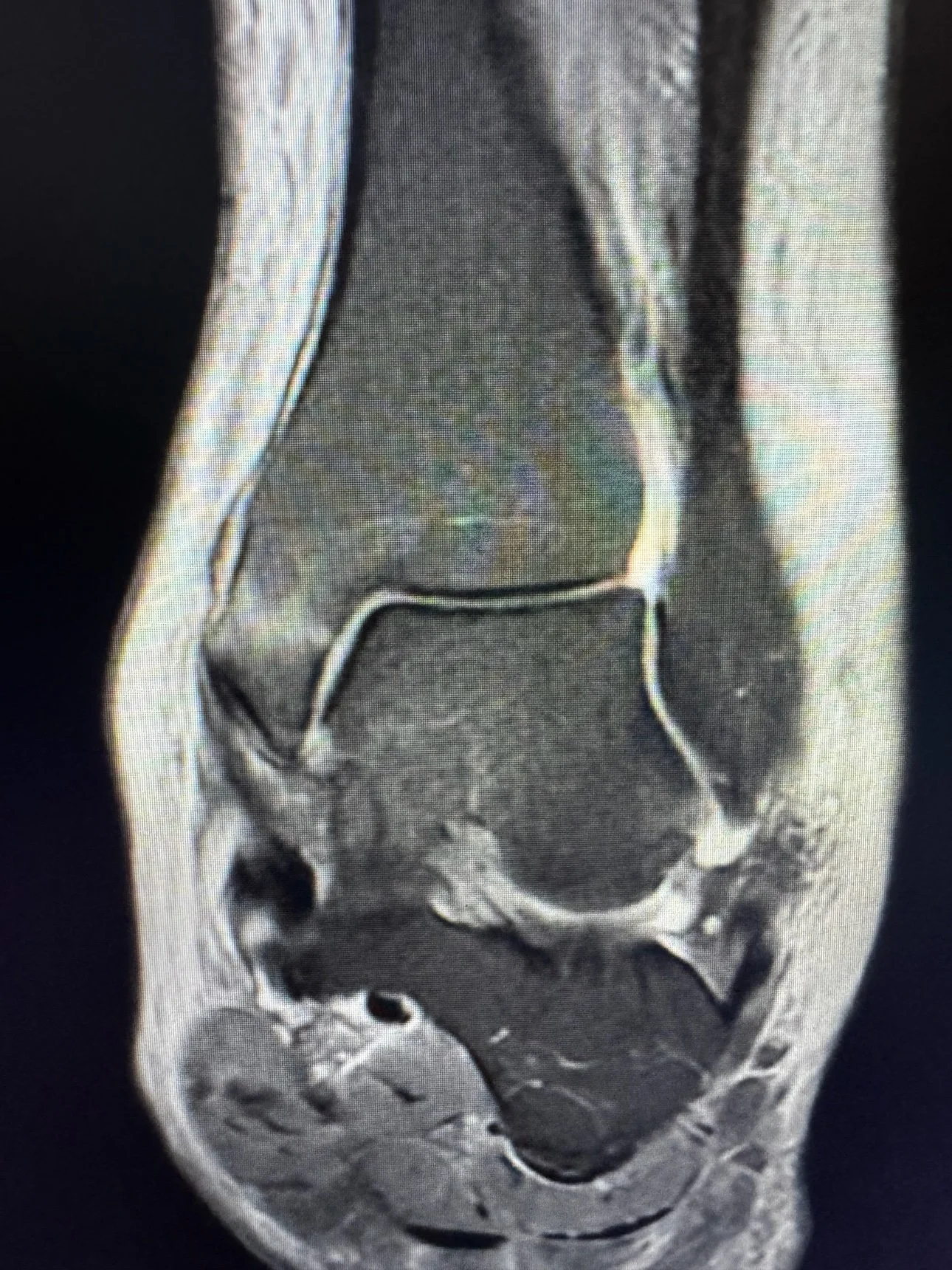
The quiet version, on MRI. A nondisplaced fracture of the medial malleolus, the inner ankle bone, with the socket still lined up. This is the pattern the bullet above describes, the one that earns a look at the whole leg and then, when the ankle is stable, heals without surgery.
The question underneath every pattern is whether the talus is still centered in the socket. A centered talus under a stable fracture can heal in a cast. A shifted talus grinds the joint cartilage unevenly with every step, and that is the setup for arthritis. Most decisions in this guide come back to that one picture.
🔗 Related Reading: Foot and Ankle Anatomy: Bones, Joints, Ligaments & Tendons Explained: The plain language tour of the bones and ligaments named here, if you want the map before the repair.
The ER and the Imaging
The first X-ray answers whether a bone is broken and whether the joint is lined up.
A stress X-ray, taken while gentle pressure twists the ankle, shows whether a fracture that looks stable is actually loose. That distinction, stable or unstable, separates the cast from the operating room for outer bone fractures.
A CT scan shows the back of the joint, where the posterior malleolus fragment hides on a plain X-ray. The CT maps its size, its shape, and whether it carries the ligament attachment that holds the fibula in place. When a posterior fragment is present, I plan the operation from the CT, not the X-ray.
A badly swollen ankle sometimes cannot be operated on safely the same day, because closing skin over that much swelling invites wound problems. The standard move is a splint, elevation, and a short wait until the skin wrinkles again when pinched gently. Waiting a few days for the skin is not a delay in your care. It is part of doing the operation well.
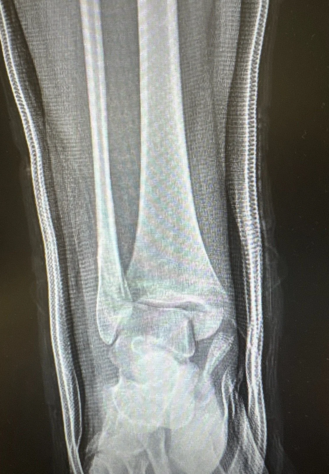
After a reduction and a splint, this ankle is still not lined up. Both ankle bones are broken, the ligaments between the leg bones are torn, and the talus still sits shifted to the outside. A splint can hold position. It cannot fix this pattern, and this is the picture that sends an ankle to surgery.
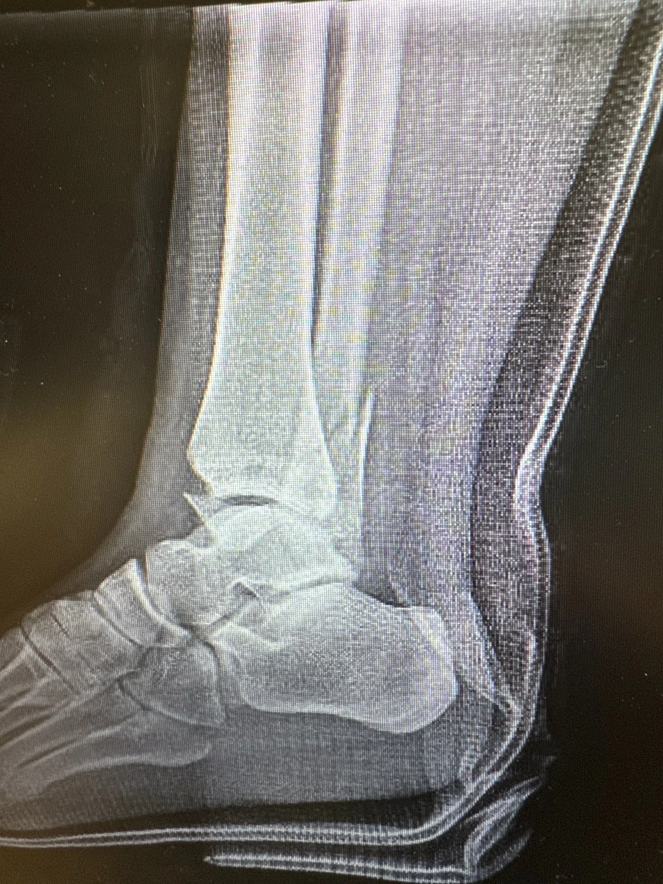
The same ankle from the side, in its splint.
Surgery or Not
Most ankle fractures that reach a surgeon are the unstable kind, and unstable ankles need surgery. The exception is narrow: the fracture is in the outer bone only, the talus is centered, and the stress X-ray shows the joint holds together under pressure. That pattern is called a stable Weber B fracture, named for where the fibula breaks relative to the syndesmosis.
In a randomized trial of 247 patients, 3 weeks in a cast or a removable boot healed stable Weber B fractures just as well as 6 weeks in a cast.¹ The boot group moved the ankle more freely and had fewer leg blood clots.
Everything outside that narrow picture, a shifted talus, two or three broken walls, a torn syndesmosis, a fracture that moves on the stress view, gets surgery. The reason is not the broken bone. Bones are good at healing. The reason is the joint surface, because a socket that heals even a couple of millimeters off wears its cartilage out years early.
🔗 Related Reading: Ankle Sprain Treatment Guide: Rehab, Braces & Recovery Timeline: A sprain is the ligament version of this injury with no broken bone, and this guide covers how those heal and how to tell the two apart.
The Operation
Ankle fracture surgery is called ORIF, short for open reduction and internal fixation. Open reduction means the surgeon lines the bones back up where they belong. Internal fixation means plates, screws, or a rod hold them there while they heal. Most patients go home the same day or after one night.
The standard construct for the outer bone is a plate along the fibula with screws. The inner ankle usually takes two screws. What gets decided case by case is the outer bone construct, plate or nail, and what happens at the back of the ankle. Those are the next two sections.
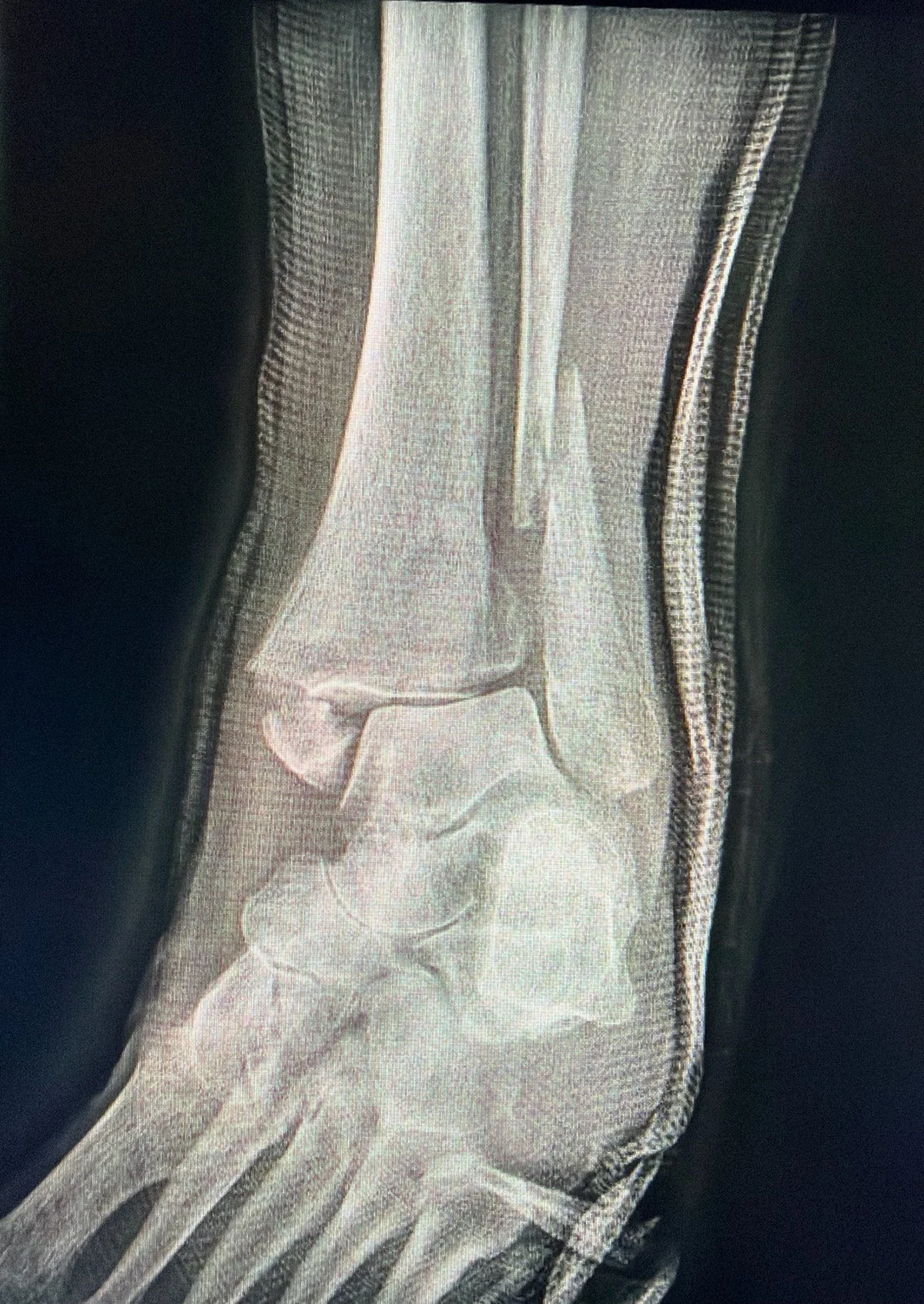
The same ankle, before. A trimalleolar fracture in its splint with the joint shifted out of the socket.
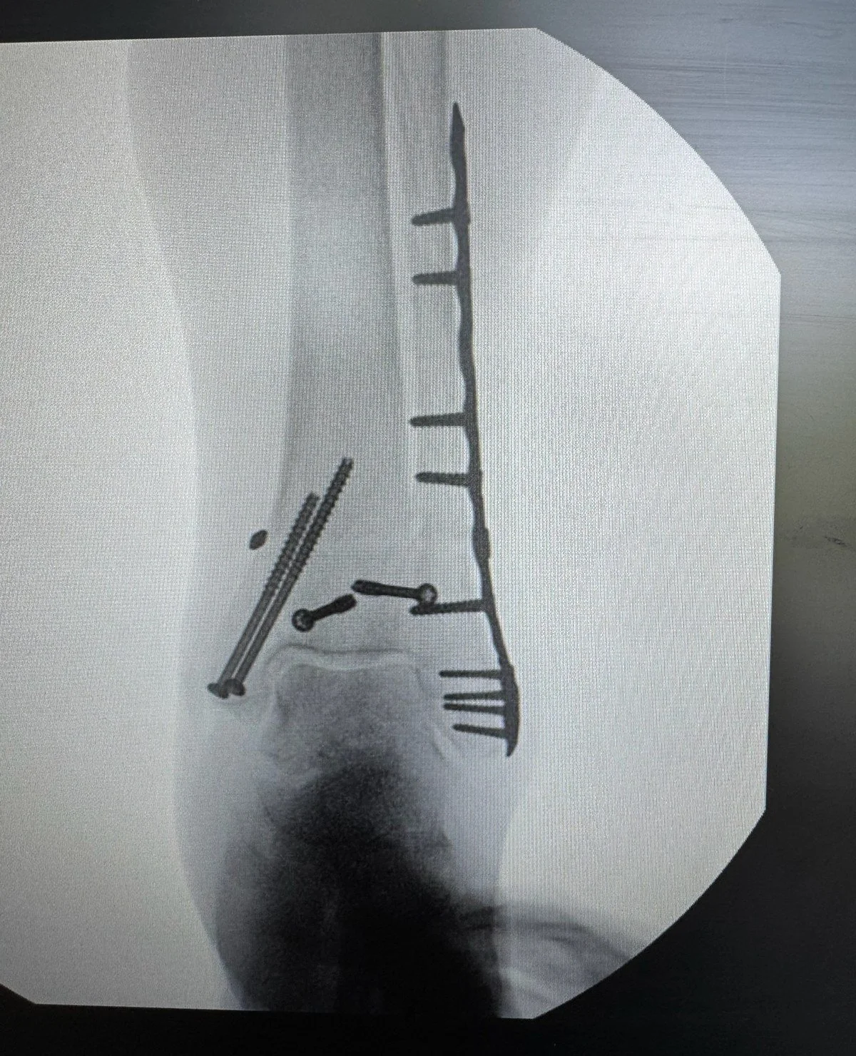
After surgery. A plate on the fibula, screws in the medial malleolus, and syndesmotic fixation, with the talus centered back in the socket. This pair of films is one case, before and after.
The Fibular Nail: A Rod Instead of a Plate
A fibular nail is a slim metal rod that slides up the center of the fibula from its tip and locks in place with small screws through its top end. Engineers call that load sharing. The rod and the bone carry the load together. Where a plate sits on the surface of the bone under a long incision, the nail works from inside the bone through an incision about an inch long.
I reach for the nail in a few situations. Unstable outer bone fractures where the pattern suits a rod. Older patients, whose skin tears if you look at it wrong and whose wounds forgive nothing. Anyone whose soft tissue worries me, including many with diabetes, where a smaller incision matters most. The worse the skin and swelling, the more the nail earns its place.
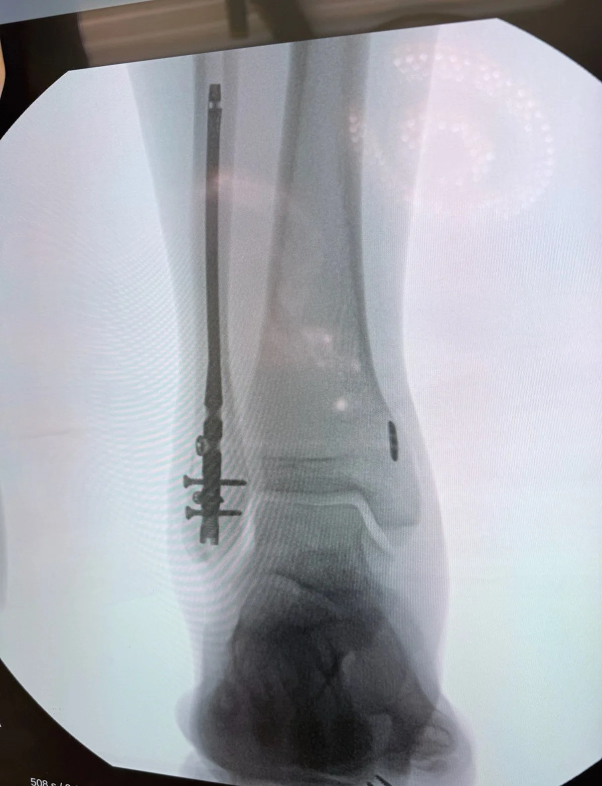
From my operating room. A fibular rod inside the outer bone, with flexible syndesmotic fixation. The button of the device sits on the inner tibia. Small incisions, hardware inside the bone, nothing prominent under the skin.
The best test of the nail is a randomized trial of 125 patients under 65 with unstable ankle fractures, nail against plate. At one year the scores were effectively tied, 78.4 for the nail and 80.2 for the plate, and stayed tied out to two years, with complications and reoperations matched. The one visible separation was wound infections, 2 with the nail against 9 with plates, a difference that fits the smaller incision though the trial was not big enough to prove it. The authors make the same point I make to patients. In younger patients the wound advantage is real but modest. In older patients it is the whole argument.²
A meta-analysis of 7 trials and 586 patients found no difference in ankle function at 3, 6, or 12 months, infections about a quarter as often with the nail, and hardware failure about twice as often.³
A single center comparison of the two constructs found complications in 23% of nail patients against 45% of plate patients, with the nail group healing about 2 weeks faster and hardware irritation a recurring complaint on the plate side.⁴
A nail demands the same respect for alignment as a plate. It just pays for that alignment with a much smaller wound.
The Back of the Ankle: The Posterior Malleolus
The posterior malleolus is the back lip of the tibia, and it does two jobs. It forms part of the joint surface your talus rolls against, and it anchors the PITFL, the posterior inferior tibiofibular ligament, the back strap of the syndesmosis. The fibula sits in a groove on the back of the tibia called the incisura, and fixing that fragment pulls the fibula back into its groove and tightens the whole socket. A posterior fragment is not just an extra piece of broken bone. It is a piece of the ligament system, in bone form.
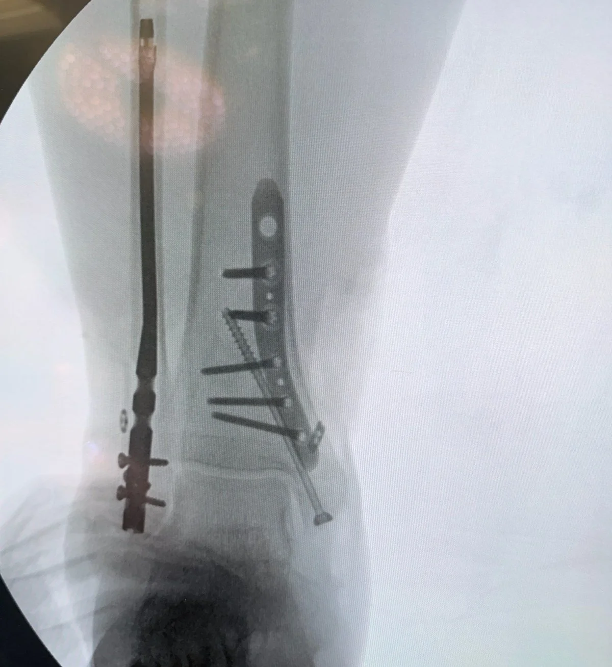
A trimalleolar fracture from my operating room, fixed with all three decisions in one ankle. A fibular nail in the outer bone, a posteromedial plate on the back fragment because that is where the fragment lived, and a lag screw in the medial malleolus.
For decades the decision ran on a ruler. Fix the fragment if it made up more than 25% of the joint surface, leave it if it did not. That rule is dead, and the CT scan killed it. Fragment shape matters more than fragment size, because a small fragment carrying the ligament attachment, sitting in the incisura, can matter more than a larger piece of the back rim that carries nothing. A 2019 case series of 50 patients planned every operation from the CT using a shape based classification, fixing fragments through the approach their anatomy called for and using syndesmotic fixation alone for the type that is really a ligament injury with a fleck of bone. Their patients scored about 74 on the standard ankle score. It is one series, not a randomized trial, but the logic is the logic I operate by. Plan from the CT, fix the fragment its anatomy calls for.⁶
When the fragment needs fixing, I fix it directly from the back, and the approach depends on where the fragment lives.
- Posterolateral approach. The incision sits at the back and outer side of the ankle and reaches the common fragment, on the back outer corner. The fragment is lined up under direct vision and a buttress plate laid against its back surface. Buttress means the plate works like a doorstop, blocking the fragment from sliding up and back.
- Posteromedial approach. The incision sits at the back and inner side. Some fragments extend toward the inner ankle or split off a separate posteromedial piece, which the outer window cannot reach or hold properly. The plate goes on through the medial window, across the fragment it was built for.
| What the CT shows | Approach | Fixation |
|---|---|---|
| A fragment on the back outer corner, carrying the ligament attachment | Posterolateral | A buttress plate against its back surface |
| A fragment extending toward the inner ankle, or a separate posteromedial piece | Posteromedial | A plate through the medial window |
| A ligament injury with a fleck of bone | No direct approach | Syndesmotic fixation alone |
Fixing the fragment directly beats the older alternative, driving screws from front to back and hoping they caught it. A systematic review found ankle scores of about 91 after direct fixation against about 83 with front to back screws, and about 92 for fixing against about 82 for leaving it alone in the studies with an untreated comparison.⁷ A meta-analysis of plate against screws found the plate left the joint surface smoother, with less step-off and gap, while scores, arthritis rates, infections, and complications came out the same.⁸ My read is simple. When the fragment is big enough to plate, the plate buys the smoother joint, and the smoother joint is what patients live with for the next thirty years.
The Syndesmosis: The Ligaments Between the Bones
The syndesmosis is the set of strong ligaments binding the fibula to the tibia just above the ankle joint. Tear it and the socket splays. Two options hold it while it heals, a screw across both bones or a suture button, a pair of small metal buttons connected by strong suture that bridges the bones like a tightrope.
The longest randomized comparison followed 43 patients with the severe rotational fracture pattern for at least 6 years, averaging 7. Both methods held the reduction equally well. Ankle scores averaged 88 with screws and 78 with the suture button, a gap that was not statistically significant in a trial this size, and arthritis a grade or more worse than the uninjured side was common in both groups.⁹ At long follow up, the two are closer than the marketing on either side suggests.
The practical differences live in the first year. A screw is rigid. It can break or loosen, and taking it out means a second operation. A suture button flexes slightly the way the natural syndesmosis does, is not designed to come out, and tolerates earlier weightbearing better. When I use flexible fixation, as in the nail film above, that forgiveness is part of the reason.
A Camera Inside the Joint: Arthroscopy During Fracture Surgery
An X-ray shows bone. It does not show the cartilage surface the bone carries, and a fracture that looks clean on film can hide real damage inside the joint. Ankle arthroscopy puts a small camera into the joint during the fracture operation, and what it finds changes the picture often enough to matter.
Across studies that scoped ankles during fracture fixation, cartilage and osteochondral injuries, damage that reaches from the cartilage into the bone under it, turned up in 27% to 89% of patients, with one systematic review averaging about 55%. The camera also finds loose fragments, ligament tears, and syndesmosis problems fluoroscopy can call normal when they are not. It checks the reduction from inside, the one view that shows whether the joint surface is flush.¹⁰
| What the camera finds | How often, or why it matters |
|---|---|
| Cartilage and osteochondral injuries | 27% to 89% of fractured ankles across studies, about 55% on average |
| Loose fragments, ligament tears, syndesmosis problems | Present even when fluoroscopy reads them as normal |
| The reduction itself | Checked from inside, the one view that shows whether the joint surface is flush |
Finding damage is only worth something if patients do better. A meta-analysis of 10 comparative studies and 755 patients found arthroscopy assisted fixation produced better function scores and lower pain scores than standard fixation, with no difference in complications or operating time.¹¹ The same review that produced the detection figures above found the same direction in the comparative studies it pooled.¹⁰ For the posterior fragment, a review of 7 studies and 101 patients found the same pattern, with direct vision of the fragment its clearest value.¹²
Most of this evidence is arthroscopy finding things, plus small, mostly retrospective comparative studies that pool different fracture types and techniques. No large randomized trial has proven that adding the camera makes ankles last longer or keeps arthritis away. What is proven is that the damage is there and imaging misses it. What is likely, on early comparative data, is that treating it helps function and pain. What is not yet proven is the long term payoff. I use the scope in fracture cases to check on the surface of the joints, to make sure no large defects are missed and that the cartilage surfaces are well aligned with no step off, and I tell patients exactly that balance.
Recovery and Weightbearing
Bone healing runs on a fairly reliable clock. Most ankle fractures are healed enough to leave the boot behind at about 6 to 8 weeks. Recovery, meaning motion, strength, balance, and trust in the ankle, runs longer, usually 3 to 6 months, and traces of swelling can hang around for the better part of a year.
For fibular nails with otherwise stable patterns I regularly allow immediate boot weightbearing. The incisions are small, and keeping an otherwise healthy person off a leg for 6 weeks costs muscle, balance, and morale they then have to buy back. Stable in that sentence has a specific meaning: the nail construct is secure, no screw crosses the syndesmosis, and a posterior fragment is either fixed solidly or does not need fixing.
| Your fixation | When walking in the boot starts |
|---|---|
| Fibular nail in a stable construct | Right away |
| Plate, well fixed | About 2 weeks |
| Syndesmosis held with a rigid screw, or a posterior fragment with tenuous fixation | Slower, on your surgeon's timeline |
| Fragile bone, or diabetes with reduced feeling in the foot | Slower, with closer follow up |
The 2 week row is not a guess. The WAX trial randomized 561 adults with surgically fixed unstable ankle fractures to start walking at 2 weeks or at 6 weeks, and walking at 2 weeks was non-inferior, the trial's term for no worse, on ankle function at 4 months.¹³ The geriatric nail trial makes the same point from the other side. Its patients walked sooner because the protocol allowed it, not because the implant is magic.⁵ Permission is the intervention, and a well fixed plate in the right pattern earns the same permission on a slower ramp. Your surgeon's instructions override anything written here.
A typical path looks like this.
| When | What happens |
|---|---|
| Weeks 0 to 2 | Splint, then a boot. Elevation above heart level as much of the day as you can. This is the swelling fight, and it matters more than anything you do later. |
| Weeks 2 to 6 | Walking in the boot if your pattern and fixation allow it. Sutures out, first X-rays, gentle motion if your surgeon clears it. |
| Weeks 6 to 8 | X-rays to confirm healing, then weaning out of the boot into a supportive shoe, often with a lace-up brace. |
| Months 2 to 6 | Physical therapy for motion and strength, balance work, return to impact and sport in stages once strength and motion earn it. |
Rehab and Stiffness
The operation fixes the bone. Rehab decides what the ankle is worth afterward. The enemy is stiffness, particularly losing dorsiflexion, the ability to pull the foot up toward your shin. An ankle that heals straight but barely moves has not finished the job.
Therapy starts with motion, then strengthening, then balance and proprioception, your joint's sense of where it is in space, the sense that keeps you from rolling the ankle again. The early weightbearing trials above doubled as rehab trials. The groups that moved and loaded early had better motion and function at 6 weeks. Motion is not the reward for healing. It is part of the healing.
The mistake I see most is too much motion too soon. The pain starts improving, and they start ignoring weight bearing restrictions or going without the boot too soon.
Risks and Complications
Arthritis. This is the long shadow of any fracture that enters a joint. Anatomic reduction, getting the joint surface flush, is the surgeon's main tool against it, and arthritis a grade or more worse than the uninjured side was common in the long term syndesmosis trial anyway.⁹ Arthritis risk is real. It is not a certainty, and many people with a stiff X-ray have a comfortable ankle.
🔗 Related Reading: Ankle Arthritis: Symptoms, Causes & Best Treatment Options: What post-traumatic arthritis feels like years later, and the full range of what can be done about it, surgical and not.
Hardware irritation. Plates sit under the skin where shoes and boots rub. In the studies above it was the plate group's recurring complaint and the leading reason for a second operation to remove hardware.⁴˒¹⁴ Hardware that bothers you after the bone has healed can usually come out in a short outpatient procedure.
Wounds and infection. The risk climbs with swelling at the time of surgery, smoking, and diabetes. It is why surgery sometimes waits for the wrinkle sign, and why small incision techniques earn their keep in fragile skin.
Slower healing with diabetes. Fractures in patients with diabetes, particularly with reduced feeling, heal slower and fail in ways other ankles do not, including collapse of the healing bone into a Charcot process. Those ankles get longer protection, closer X-ray follow up, and a lower threshold to call.
🔗 Related Reading: Charcot Foot: The Diabetic Complication That Changes Everything: Why a fracture or injury in a numb diabetic foot is a different emergency, and how the collapse happens.
Stiffness, blood clots, and nerve irritation round out the list. Stiffness is common and usually yields to therapy. Blood clots are uncommon after ankle surgery but real, and calf pain or new swelling after the first week is a call, not a wait. Numbness around an incision usually fades over months.
When to Call
Call the office, or go in, for any of these.
- Fever over 101, spreading redness, or drainage from the incision
- Pain that is getting worse after it had been getting better
- Toes that turn pale, blue, or cold, or swelling that makes the splint or boot feel tight enough to cut in
- Calf pain or new calf swelling
- A fall onto the ankle, or numbness that is spreading rather than shrinking
❓ Frequently Asked Questions
How long does a broken ankle take to heal?
The bone itself usually heals in 6 to 8 weeks. Most people are in a regular shoe by about 2 months, with motion and strength improving for 3 to 6 months. Some swelling on a long day can persist toward a year.
Does every broken ankle need surgery?
No. A stable fracture, outer bone only, with the talus centered and the joint holding together on a stress X-ray, heals in a cast or boot, and trial data says 3 weeks of immobilization can be enough for that pattern.¹ Surgery is for unstable ankles, where the joint would heal out of position.
When can I walk on it after surgery?
For fibular nails with otherwise stable patterns I regularly allow immediate boot weightbearing. Stable there means secure fixation, no screw across the syndesmosis, and a solid posterior fragment. Many plate patients start walking at about 2 weeks, which the WAX trial showed is no worse than waiting 6.¹³ Syndesmosis screws and certain posterior fragments still earn a slower timeline.
Will I get arthritis from this?
Your risk is higher than it was, and it is not a promise. Arthritis a grade or more worse than the uninjured side was common in the long term syndesmosis trial at 7 years, and many scored their ankles well anyway.⁹ Getting the joint surface flush at surgery is the main protection.
Does the hardware have to come out?
Usually not. Hardware comes out when it causes trouble, most often a plate rubbing under the skin. The exception is a syndesmosis screw, sometimes removed in a planned second operation after the ligaments heal. A nail inside the bone rarely bothers anyone.
Why a nail instead of a plate for the outer bone?
Same job, different address. The plate sits on the bone's surface under a longer incision. The nail sits inside the bone through a small incision and shares load with it. Trials show the same function at a year, fewer wound infections trending with the nail, and less hardware irritation.²˒⁴ For older patients and fragile skin, that trade is usually worth it.
Products I Recommend for Ankle Fracture Recovery
Three things earn their keep after a broken ankle. Everything else is optional.
- CAM Walker Boot: The boot is the cast you can take off to wash and, when cleared, to move. Get the fit right. You will live in it for weeks.
- EvenUp Walking Boot Leveler: A boot sole is thick. This levels the other shoe to match, so your hips and back are not paying for your ankle's recovery.
- Ankle Ice Pack: Cold and elevation are the swelling program for the first two weeks. A pack shaped for the ankle gets used. A bag of peas gets forgotten.
Note: These are general recommendations I use in practice. Some links are affiliate links which support Feet Made Simple at no cost to you. Always confirm with your provider before starting new treatments.
References
- Kortekangas T, Haapasalo H, Flinkkilä T, Ohtonen P, Nortunen S, Laine HJ, Järvinen TLN, Pakarinen H. Three week versus six week immobilisation for stable Weber B type ankle fractures: randomised, multicentre, non-inferiority clinical trial. The BMJ. 2019;364:k5432. DOI: 10.1136/bmj.k5432. PMID: 30674451.
- White TO, Bugler KE, Olsen L, Lundholm LH, Holck K, Madsen BL, Duckworth AD. A prospective, randomized, controlled, two-center, international trial comparing the fibular nail with open reduction and internal fixation for unstable ankle fractures in younger patients. Journal of Orthopaedic Trauma. 2022;36(1):36-42. DOI: 10.1097/BOT.0000000000002140. PMID: 33878069.
- Zhang J, et al. The fibular intramedullary nail versus plate fixation for ankle fractures in adults: a systematic review and meta-analysis of randomized controlled trials. Journal of Orthopaedic Surgery and Research. 2024;19:542. DOI: 10.1186/s13018-024-05032-z. PMID: 39238000.
- Rushing CJ. Comparison of ankle fracture fixation using intramedullary fibular nailing versus plate fixation. The Journal of Foot & Ankle Surgery. 2024;63(5):546-556. DOI: 10.1053/j.jfas.2024.05.004. PMID: 38825307.
- Kohler FC, Berfelde Z, Schenk P, Weschenfelder W, Wildemann B, Kobbe P, Mendel T, Ullrich BW, et al. Fixing geriatric ankle fractures: fibular nail versus locking plate in a prospective multicenter study. European Journal of Trauma and Emergency Surgery. 2026;52(1):52. DOI: 10.1007/s00068-026-03108-5. PMID: 41729233.
- Mason LW, Kaye A, Widnall J, Redfern J, Molloy A. Posterior malleolar ankle fractures: an effort at improving outcomes. JBJS Open Access. 2019;4(2):e0058. DOI: 10.2106/JBJS.OA.18.00058. PMID: 31334465.
- Miksch RC, et al. Open reduction and internal fixation of the posterior malleolus fragment in ankle fractures improves the patient-rated outcome: a systematic review. Foot & Ankle International. 2023;44(8):727-737. DOI: 10.1177/10711007231165771. PMID: 37125766.
- Wang J, Jia HB, Zhao JG, Wang J, Zeng XT. Plate versus screws fixation for the posterior malleolar fragment in trimalleolar ankle fractures. Injury. 2023;54(2):761-767. DOI: 10.1016/j.injury.2022.10.032. PMID: 36336477.
- Lehtola R, Leskelä HV, Flinkkilä T, Pakarinen H, Niinimäki J, Savola O, Ohtonen P, Kortekangas T. Suture button versus syndesmosis screw fixation in pronation-external rotation ankle fractures: a minimum 6-year follow-up of a randomised controlled trial. Injury. 2021;52(10):3143-3149. DOI: 10.1016/j.injury.2021.06.025. PMID: 34246483.
- Meyer-Pries M, Hajymiri M, Lytras T, Manolopoulos P, Ntourakis D. Arthroscopy-assisted open reduction internal fixation versus conventional open reduction internal fixation in the treatment of ankle fractures: a systematic review with meta-analysis. HSS Journal. 2025;21(1):86-92. DOI: 10.1177/15563316231204616. PMID: 39846056.
- Zhuang C, Guo W, Chen W, Pan Y, Zhuang R. Arthroscopically assisted internal fixation for treatment of acute ankle fracture: a systematic review and meta-analysis of comparative studies. PLoS ONE. 2023;18(8):e0289554. DOI: 10.1371/journal.pone.0289554. PMID: 37540648.
- MacFarlane EM, Qureshi A, Boggiano VJ, Kennedy JG, Gianakos AL. Arthroscopic assisted fixation of posterior malleolus fractures: technical pearls and clinical outcomes. Foot and Ankle Surgery. 2026;32(3):238-246. DOI: 10.1016/j.fas.2025.09.003. PMID: 40987675.
- Bretherton CP, Achten J, Jogarah V, et al.; WAX Investigators. Early versus delayed weight-bearing following operatively treated ankle fracture (WAX): a non-inferiority, multicentre, randomised controlled trial. The Lancet. 2024;403(10446):2787-2797. DOI: 10.1016/S0140-6736(24)00710-4. PMID: 38848738.
- Dehghan N, McKee MD, Jenkinson RJ, Schemitsch EH, Stas V, Nauth A, Hall JA, Stephen DJ, Kreder HJ. Early weightbearing and range of motion versus non-weightbearing and immobilization after open reduction and internal fixation of unstable ankle fractures: a randomized controlled trial. Journal of Orthopaedic Trauma. 2016;30(7):345-352. DOI: 10.1097/BOT.0000000000000572. PMID: 27045369.
- Lokerman RD, Smeeing DPJ, Hietbrink F, van Heijl M, Houwert RM. Treatment of a scientifically neglected ankle injury: the isolated medial malleolar fracture. A systematic review. The Journal of Foot & Ankle Surgery. 2019;58(5):959-968. DOI: 10.1053/j.jfas.2018.12.028. PMID: 31178394.