
Updated by Dr. Dawson November 2026
You buy wider shoes. You loosen the laces. You start choosing restaurants based on how far you have to walk from the parking lot.
At some point, that bump beside your big toe starts making more decisions than it should.
The good news is that having a bunion does not automatically mean you need surgery. There are practical ways to make your foot more comfortable, and several effective surgical options when those changes are no longer enough.¹
TL;DR
A bunion is a change in bone alignment, not simply an extra lump of bone.¹
Shoes, padding, and sometimes inserts or spacers can help with discomfort. They do not reliably straighten an established bunion.¹ ²
Surgery is worth discussing when pain keeps interfering with life despite reasonable nonsurgical treatment.¹
Modern surgery includes both minimally invasive and open techniques. The best choice depends on your foot and the problem being corrected.³ ⁴
Walking after surgery and being fully recovered are different milestones. Some patients can walk in protective footwear right away, but healing still takes time.⁵
What exactly is a bunion?
A bunion forms when the big toe leans toward the second toe and the long bone behind it shifts out of alignment. The joint then sticks out along the inside of the foot. The medical name is hallux valgus.¹
Think of a cabinet door hanging crooked. The corner sticking out is what you notice, but sanding that corner down does not fix the way the door hangs.
That is why correcting a bunion usually involves more than shaving off the bump.
Why did I get one?
Usually, several factors are involved. The foot structure you inherited, how your joints move, and the shoes you wear can all play a part.⁶
Tight shoes can squeeze the area and make symptoms worse. But plenty of people with bunions have never spent a day in high heels.
So, no, this is not necessarily punishment for your footwear choices in your 20s.
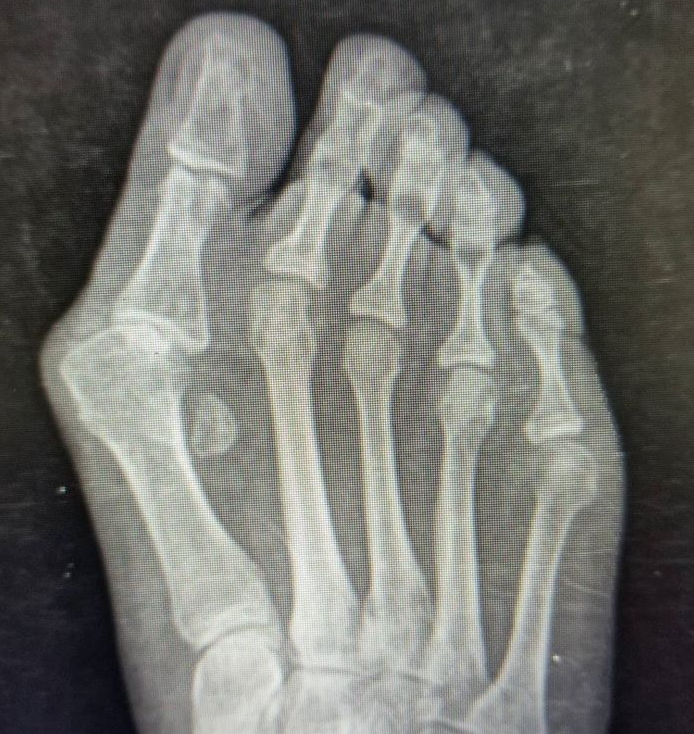
This condition is not just cosmetic. It is associated with pain, decreased walking efficiency, increased fall risk in elderly patients, and reduced quality of life.³
Common companions: Pain under the big toe joint is often sesamoiditis. Numbness between the lesser toes can be Morton’s neuroma.
What helps, and what it can actually do
Roomier shoes + thin padding
Less squeezing and rubbing. The bunion itself remains.¹
Inserts
May improve comfort and pressure distribution. They do not permanently straighten the bones.²
Spacers + splints
May help symptoms. Lasting correction is not reliably established.²
Surgery
Can correct alignment when symptoms justify it. Requires healing time and carries risks.¹
Shoe comfort and mechanics: If arch or heel pain limits your walks, compare with plantar fasciitis. Nail edge tenderness from crowding fits the pattern in ingrown toenails.
What the footwear evidence actually says. In a 2024 gait laboratory study of 28 women with painful bunions, extra width shoes fitted with prefabricated arch contouring orthoses cut the peak pressure against the inside of the big toe joint from 58.3 to 42.6 kilopascals compared with the women's own shoes.¹⁴ Pressure is the mechanism, not the whole story: whether that pressure drop turns into less pain is exactly what the study could not prove, and a systematic review of nonsurgical care rates the overall evidence for symptom relief as limited.² So the honest claim is a narrower one. Roomier shoes unload the joint. That is a real effect, and it is not the same as a cure.
The features matter more than the label. A wide toe box that does not squeeze the forefoot, a midfoot that holds the shoe on without laces doing the squeezing, and enough cushion for your walking surface. Any brand that delivers those 3 things is doing its job. The specific models I point patients to are in the products section at the end of this article, labeled as my picks, because that is what they are.
2 supporting tools worth knowing. An insert with a Morton's extension (a small build up under the big toe joint) can protect the joint and shift load in feet where flatfoot or forefoot pain travels with the bunion. And plain anti inflammatory basics still count: an NSAID when you tolerate it, ice after a long day. Neither changes the alignment. Both can buy comfort while you decide what the foot actually needs.
When is it time to get your bunion checked?
Make an appointment if pain keeps returning, reasonable shoes still hurt, or you are changing your activities because of your foot.¹
An evaluation should include a conversation about where it hurts and what you want to get back to. An examination and standing X-rays help show the alignment and whether the joint itself has other problems.³ ⁵
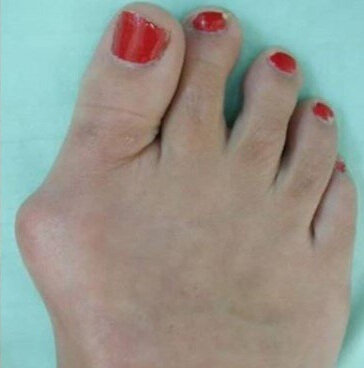
A photo shows the bump. It does not tell the whole story.
How do you know when surgery is worth it?
A useful question is: “Am I managing this comfortably, or am I organizing my life around it?”
Surgery becomes worth discussing when appropriate nonsurgical treatment has not provided enough relief. Difficulty walking, exercising, working, or finding comfortable shoes can all be part of that conversation.¹
You do not have to wait until the toe is dramatically crooked. You also do not need surgery simply because the bump is visible.
The purpose is to improve comfort and function. Operating only to change how a foot looks exposes you to risks without the same functional reason for treatment.⁷
What does bunion surgery actually do?
Most operations work by changing the position of the bones and balancing the tissues around the joint. The correction is often held with screws, a plate, or another implant while it heals.⁵ ⁸
There are several ways to do that. The names can sound like a menu written in another language, but the basic choices are easier to understand.
Bone realignment: moving the pieces into a better position
An osteotomy is a controlled cut in a bone. The surgeon repositions it and secures it while it heals.
Depending on the correction needed, the cut may be near the end of the metatarsal, farther along the bone, or combined with a small correction in the big toe itself. You may hear names such as chevron, scarf, transverse osteotomy, or Akin.⁹
The important question is what the procedure will accomplish for your foot.
Lapidus: correcting through a joint farther back
A Lapidus procedure realigns and fuses the joint at the base of the first metatarsal, farther back than the visible bunion. It can be useful when that joint and the surrounding alignment need to be addressed.⁸
It does not fuse the big toe joint. Those are different joints in different places.
Minimally invasive Lapidus: same fusion, smaller doorway
The Lapidus fusion can also be done through small incisions, using a purpose built screw system to hold the correction. The system I use for it is the PROstep MIS Lapidus (Stryker). It is hardware, not a separate operation, and it is not the technique this article leads with anymore.
Read any claim about it with the calendar in hand. The system was released in 2023, and there are no published outcome studies of this specific hardware yet. What exists is early evidence for the technique as a class. A 2025 systematic review of minimally invasive Lapidus surgery found 5 studies covering 162 feet, with a total complication rate of 12.3%. The most common problems were painful hardware, nonunion (the fusion not healing solid), and the deformity coming back.¹² That is a reasonable early safety signal. It is not proof that the small incision version beats the open one, and a surgeon who tells you otherwise is ahead of the data. It stays a Lapidus: smaller doorway, same fusion to heal.
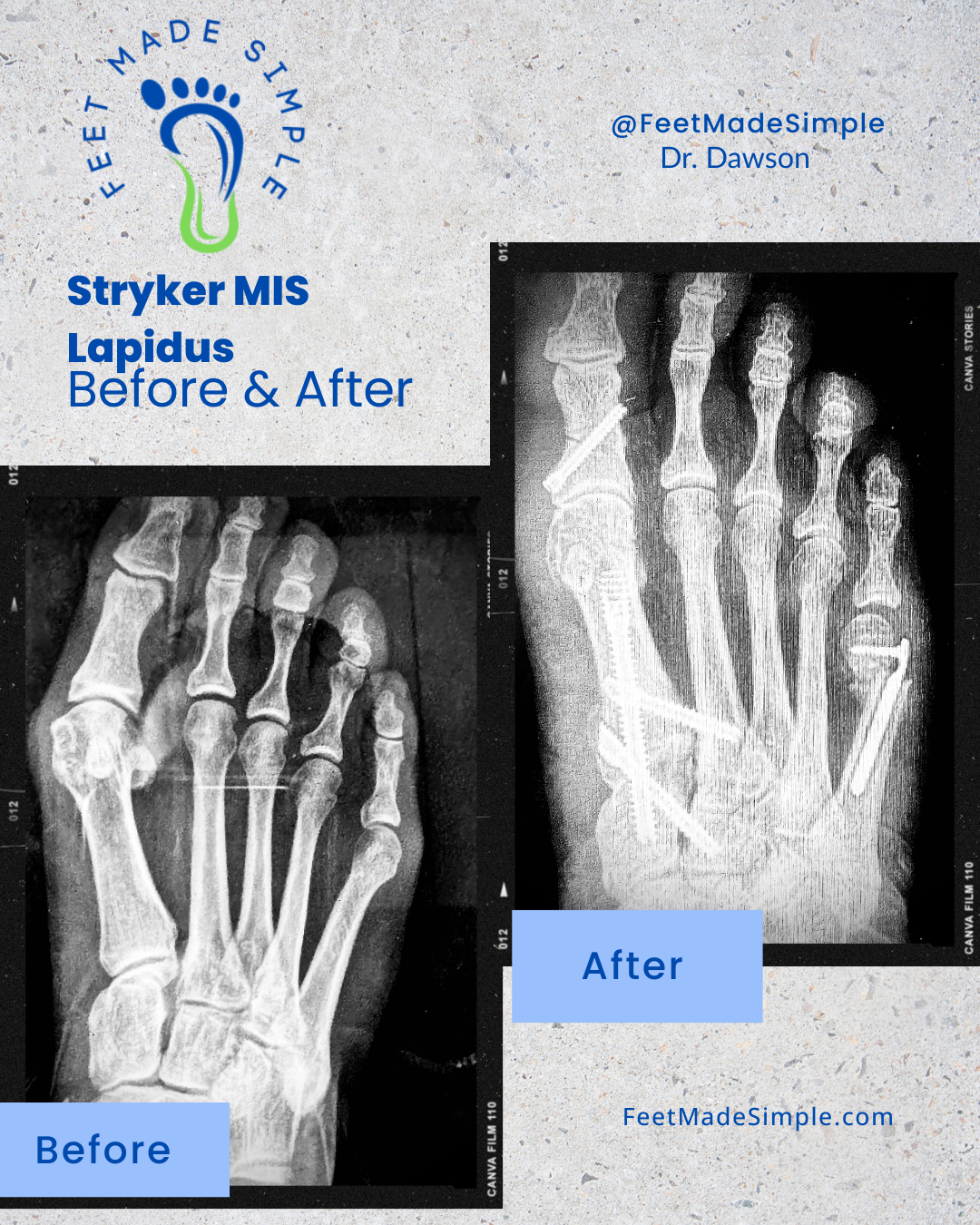
PROstep MIS Lapidus hardware (Stryker), shown with the system’s tailor’s bunion screw.
This patient was allowed to bear weight on the foot the day of surgery in a walking boot
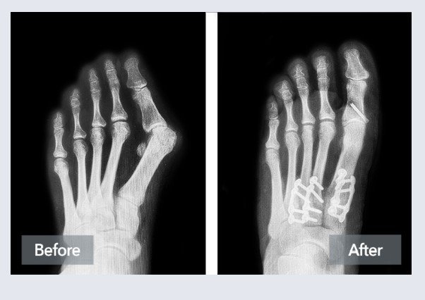
Before and after a Lapidus correction of a severe bunion using the Lapiplasty system (Treace). This variant is called Adductoplasty. It also repositions the midfoot bones next to the fused joint, which is why it is reserved for severe deformities.
Big toe joint fusion: an option for a damaged joint
If the big toe joint has substantial arthritis or certain other problems, straightening the bones alone may not address the pain. A fusion can be an appropriate option in selected cases.³ ⁹
That does mean giving up movement at that joint. The tradeoff is treating a joint that has become painful or unreliable.
Bunion operations in plain English
Minimally invasive distal osteotomy: a small cut at the neck of the bone
The metatarsal is cut at the neck through small incisions, the head is slid and rotated back into line, and 2 screws hold the correction; an Akin cut in the toe bone is added when the toe needs it.¹⁵
Open osteotomy: move the bone
A controlled cut allows the surgeon to reposition the bone. Can be performed through an open or minimally invasive approach.⁴ ⁹
Lapidus: realign and fuse farther back
Lapidus is not the default bunion operation in my practice. It is the operation for the bunion whose problem lives at the base of the 1st metatarsal: severe deformity, a loose base joint, or arthritis in that joint. When the base joint is sound, the distal osteotomy above does the job through a smaller doorway. The big toe joint remains separate.⁸
Big toe joint fusion: treat a damaged joint
Can be appropriate for significant arthritis or certain complex problems. The fused joint no longer bends.³ ⁹
Small incision or larger incision?
That describes how the surgeon reaches the bones. It is a separate question from what correction your foot needs.
Minimally invasive distal osteotomy: a small cut at the neck of the bone, a real correction
Most bunion operations move the head of the 1st metatarsal back toward the midline. This one does it through incisions you could cover with a thumbnail.
Through small incisions, I cut the 1st metatarsal at its neck with a burr, slide the head sideways, rotate it when the toe has rolled, and hold it with 2 screws. A small wedge cut in the toe bone itself, the Akin, finishes the straightening when the toe needs it. The operation comes out of the Vernois and Redfern school of percutaneous forefoot surgery, and the modern fixed version is called MICA, short for minimally invasive chevron and Akin.¹⁵ Newer systems package the same operation. Nanoplasty (Treace) guides the cut with a small saw instead of a burr and holds the correction with locking fixation. It is a newer system, and it has no published outcome studies of its own yet. Everything below is the evidence for the operation as a class, not for any 1 brand.
My preference, labeled as preference and not as evidence: in a foot that fits this operation, this is the technique I now reach for first.
What the correction looks like. In the largest prospective series, 333 feet from the center that developed the technique, the hallux valgus angle went from 32.9 to 8.7 degrees and the angle between the 1st and 2nd metatarsals from 15.3 to 5.7 degrees at 2 years, while the pain score on the Manchester Oxford Foot Questionnaire fell from 44.5 to 9.4.¹⁶ A center that did not invent the operation reported the same shape of result in 40 patients, with the questionnaire score improving from 58 to 10 at 1 year.¹⁷ The newest version swaps the chevron cut for a straight transverse cut and corrects the rotation of the bone more deliberately; in 50 feet it produced the same order of correction at 1 year.¹⁸ Evidence grade: Moderate. Prospective data with real numbers, still concentrated in the hands of the surgeons who built the technique.
What it does not beat. Head to head, the small incision has not proven itself the better operation. It has proven itself the equal one. Against open distal chevron, outcomes and complications matched at 2 years.¹⁹ A randomized trial against open Scarf found equal scores and angles at 6 months, with less pain in the percutaneous group from day 1 through week 6.²⁰ A matched comparison found the same tie at 2 years, with shorter operations and the only wound complications on the open side.²¹ A 5 year randomized trial found no difference in anything measured.²² And the strictest pooling of the controlled studies put the score advantage for the minimally invasive side at 4 points, under the 29 points a patient can actually feel, with no difference in pain, angles, or complications. Its authors would not recommend it over open surgery on that evidence.²³ Evidence grade for better than open: not established. Evidence grade for as good as open, with an easier first few weeks: Moderate.
Recovery. The rigid fixation is the point of the modern version. In the large series, patients walked in a flat rigid sandal from day 1 and went home the same day.¹⁶ Read that correctly: walking means the bathroom, the kitchen, short trips around the house. The bone still needs about 6 weeks to heal, and normal shoes and a job on your feet wait for healing, not for the calendar of your hopes. What the small incisions buy is a quieter wound and, in the comparative studies, less pain in the first weeks.²⁰ ²¹
What can go wrong. Screws are the tax on this operation. In the 333 foot series, 6.3% of feet later had screws removed, 1.2% had a deep infection that needed surgical cleaning, 1.5% had a slow union, and 0.9% each had the deformity come back with symptoms and pain shifted under the neighboring metatarsals. That last one is transfer metatarsalgia: move the 1st metatarsal and the load sharing across the forefoot changes with it.¹⁶ A separate series put screw removal at 7.2%, its most common complication.³¹ The operation is also technique hungry, and this part matters when you choose a surgeon. The first published series from a new center reported a 14% reoperation rate across its first 106 feet and called its own learning curve steep.²⁴ Operating time and X-ray use settled after roughly 21 to 27 cases in 1 series, and in another series complications kept improving until about case 42.²⁵ ²⁶ Asking how many of these your surgeon has done is a fair question, and the literature is why. Fluoroscopy is the other trade: the percutaneous operation used about twice the intraoperative X-ray exposure of a mini open hybrid in a prospective comparison, and the measured doses stayed below occupational safety limits.²⁷
Where it stops. A cut at the neck of the bone moves the head of the bone. It cannot stabilize a loose joint at the base of the 1st metatarsal, and it cannot fix arthritis in that joint. Those feet belong to the Lapidus fusion below. Severe deformity is the honest gray zone: a systematic review of this operation in bunions with a hallux valgus angle over 40 degrees and an intermetatarsal angle over 16 degrees found good correction and high satisfaction across 676 feet, and said in the same breath that the published evidence is limited.²⁸ I read that as a selection judgment for experienced hands, not a settled indication. One more honesty note, because it cuts against a rule you will hear stated as fact: a randomized trial found that feet with a loose base joint did no worse with a distal osteotomy than with a fusion at 2 years, so "loose joint means fusion" is a judgment, not a law.²⁹ And the field's current concepts review is blunt that most of the minimally invasive evidence is still single surgeon series.³⁰
The state of play: real correction, small wounds, walking the same day, and an evidence base still catching up to the operation's popularity.
Procedure comparison
| Procedure | What it does | Fits best | Recovery pattern | Watch for |
|---|---|---|---|---|
| Minimally invasive distal 1st metatarsal osteotomy (MICA class; Nanoplasty is 1 current system) | The metatarsal is cut at the neck through small incisions, the head is slid and rotated back into line, and 2 screws hold the correction; an Akin cut in the toe bone is added when the toe needs it | Mild to moderate bunions, and selected severe ones, when the joint at the base of the 1st metatarsal is stable and not arthritic | Walking in a rigid sandal or boot from day 1; about 6 weeks for the bone to heal | Screw removal (6.3% in the largest series), recurrence, transfer pain under the next metatarsal; a real surgeon learning curve¹⁶ ²⁴ |
| Lapidus (1st tarsometatarsal fusion) | Realigns and fuses the joint at the base of the first metatarsal. It does not fuse the big toe joint. | Severe deformity, a loose (hypermobile) joint at the base of the 1st metatarsal, or arthritis in that joint. This is the operation that treats the base joint itself. A distal osteotomy cannot stabilize it and cannot fix its arthritis.²⁹ | The fusion must heal before normal loading; protected weight bearing while it heals | Nonunion (the fusion not healing solid), painful hardware, recurrence¹² |
| Distal Chevron Osteotomy | Realignment with bone cut near toe joint, fixed with screws | Mild to moderate deformity | Early weight bearing in surgical shoe | Recurrence risk, stiffness, transfer metatarsalgia |
| Scarf Osteotomy | Z-shaped cut for moderate to severe bunion, strong fixation | Moderate to severe deformity | Early mobilization in surgical shoe | Longer scar, troughing, stiffness |
| Akin Osteotomy | Wedge cut in big toe bone to correct toe alignment | Mild deformity or as adjunct | Quick recovery, short procedure | Limited correction alone, overcorrection risk |
| Proximal Osteotomy | Cut near base of metatarsal for larger correction | Moderate to severe deformity | Longer recovery, non-weight bearing phase | Delayed union, stiffness, higher complication risk |
| MTP Arthrodesis | Fusion of big toe joint | Severe deformity with arthritis | Extended recovery, no bending at big toe joint | Nonunion, adjacent joint stress, permanent stiffness |
What about minimally invasive bunion surgery?
Minimally invasive surgery, or MIS, allows the surgeon to correct the bones through smaller incisions using specialized instruments. It is still bone surgery, even if the openings in the skin are small.⁵
Think of renovating a room through a smaller doorway. The doorway changes how you get the work done. The work inside still needs to be done properly.
Some minimally invasive operations use a chevron shaped cut. Others use a transverse cut across the bone. Fixation also varies. “MIS” describes a group of techniques rather than 1 identical operation.⁴ ¹⁰
Does it work as well as traditional surgery?
Modern MIS techniques have encouraging evidence behind them.
A randomized study found comparable results between minimally invasive and open chevron surgery at 5 years.⁴
A larger study of 729 feet treated with a minimally invasive transverse osteotomy found improvements in pain, walking, and alignment at an average follow-up of 2.6 years. The reported X-ray recurrence rate was 4.5%, and the complication rate was 6.1%. Those results describe that particular study, not a guarantee for every operation.¹⁰
That makes MIS a legitimate option. It does not mean every bunion should be treated through the smallest possible incision.
A 2026 study of severe bunions also highlighted the importance of how flexible the deformity was before surgery. Both techniques studied improved alignment, but recurrence was higher in the transverse group, which contained more stiff deformities.¹¹
My perspective
I now favor minimally invasive correction for many suitable patients, often using a transverse bone cut and an implant inside the bone to hold the correction.
But the bigger conversation is about finding the operation that fits your foot. An experienced surgeon should be able to explain why they recommend a particular approach and what alternatives make sense.
Can you walk after bunion surgery?
Sometimes, yes. Some modern procedures allow protected walking in a surgical shoe or boot immediately after surgery. Others require a period of keeping weight off the foot.⁵ ⁸
In my practice, I commonly allow protected walking from day 1 after my usual MIS correction when the patient’s health and the repair support it.
That is permission to get around safely while healing. It is not a prescription for 10,000 steps.
The implant holds the correction while the bone heals. It does not make the bone instantly healed.
“I can walk” is not the same as “I'm healed”
Early protected walking
Getting around in the prescribed shoe or boot, within your surgeon's limits.
Returning to regular shoes
A separate milestone based on healing, swelling, and the operation performed.
Long walks, full work shifts, and exercise
More demanding goals that need their own recovery plan.
Ask for a plan that matches your life, not just a date when you can “walk again.”
How long does recovery take?
Recovery is a series of milestones.
Getting to the bathroom, wearing a regular shoe, standing through a workday, and going for a run all ask different things of your foot.
Early recovery: protect the repair
Plan on protective footwear, dressing care, follow-up appointments, and time to rest.
Arrange help with the jobs that keep you standing. Grocery shopping, laundry, childcare, and an enthusiastic dog do not pause just because you have had surgery.
Returning to shoes: let healing lead
Many MIS protocols use a protective shoe for approximately 6 weeks, but the timing varies. Your examination, X-rays, swelling, and any additional procedures help determine when it is appropriate to change footwear.⁵
Even once you are cleared for regular shoes, a roomy sneaker may feel good before a dress shoe does.
Returning to normal life: expect gradual progress
Swelling can continue for months. Full recovery may take 6 to 12 months, even though you can often resume useful activities much sooner.⁵
Ask about your actual routine. “When can I work?” means something different for someone at a desk than for someone standing through a 12-hour shift.
Driving also needs its own discussion, particularly after right foot surgery or while taking sedating pain medication.
What can go wrong?
Possible complications include infection, nerve irritation, stiffness, persistent pain, problems with bone healing, uncomfortable hardware, and the bunion returning. Occasionally, another procedure is needed.⁵
Smaller incisions do not make those risks disappear.
Before deciding, ask what your surgeon sees as the biggest risks for your foot, how likely they are, and how they would be managed.
What I want you to take away
You do not need to become an expert in bunion surgery to make a good decision.
Start with comfortable shoes and practical ways to reduce irritation. If your foot still limits your life, get it assessed and ask someone to explain the options in plain English.
The goal is a comfortable, useful foot that lets you get back to the things you enjoy
Bunions in kids and teens
A bunion on a growing foot is a different problem from the adult version, and it deserves its own conversation instead of adult advice shrunk down.
Juvenile hallux valgus often runs with ligament laxity and flatfoot, and the deformity can progress while growth continues. Pain is not always the first sign. What parents usually notice first is shoe fit: shoes that wear unevenly, toes starting to overlap, a visible drift of the big toe in a kid who has not complained about anything.
The management order is conservative first, and it is not close:
- Shoes with a wide toe box, so the forefoot is not squeezed while it is still forming.
- Orthotics when flatfoot is part of the picture, to manage load, not to straighten the toe.
- Activity modification when a sport is the thing that hurts.
- No splints for correction. There is no evidence they produce a lasting change in a growing foot either, and a device a teenager will not wear corrects nothing.²
Surgery is where timing does the heavy lifting. Recurrence is the most common complication after juvenile bunion surgery, it is tied to skeletal immaturity, and recent studies put it at roughly 20%.¹³ The ACFAS consensus statement covers pediatric decision making directly, and its posture matches mine: monitor, manage symptoms, and let the foot finish growing unless pain or deformity is severe enough to force the issue.³
In a growing foot, time is a treatment. Use it unless the foot forces your hand.
Frequently asked questions
Do bunion splints fix the bone?
No. They may offer comfort but do not realign the first metatarsal. Alignment changes require surgery when symptoms and function demand it.¹
Can you just shave the bump off?
Usually, shaving alone would leave the underlying alignment problem in place. It may be part of an operation, but the correction often requires repositioning bone as well.¹ ⁵
When is surgery reasonable?
When pain or shoe conflict limits life despite shoe changes, orthoses, and activity adjustments. Imaging and exam guide the procedure choice.¹
Should I have surgery before it starts hurting?
A painless bunion is not automatically a reason to operate. Symptoms, function, and individual circumstances should guide the discussion.⁷
Will shoes alone stop progression?
Shoes can improve comfort and reduce symptoms. Progression depends on anatomy, ligament laxity, and load.¹
Will I need the screws taken out?
Usually, implants can stay in place. Removal may be considered if they become painful or cause another problem. It is not an automatic second stage of recovery.⁵
Can a bunion come back?
Yes. Recurrence can happen after different types of surgery. A change on an X-ray does not always mean the pain has returned or that another operation is needed.⁵
What if my child has a bunion?
Growing feet need their own assessment. Remaining growth, symptoms, and the risk of recurrence affect treatment decisions. Adult surgical advice should not simply be applied to a child.³ (Expanded in "Bunions in kids and teens," above.)
🦶 Recommended Products for Bunions
These items are listed for education and reference only. They are not a substitute for medical advice. Please see your podiatrist for specific recommendations. I may receive a commission if you purchase through these links, at no additional cost to you.
👟 Shoes
- Topo Ultraventure 4 – Trail shoe with wide toe box to reduce bunion irritation
- Topo Atmos – Maximalist shoe with roomy forefoot
- Altra Torin 8 – Zero-drop shoe with naturally wide forefoot fit
🦾 Orthotics
- Powerstep Pinnacle Maxx – Helps control pronation contributing to bunion pressure
Research & References
Good foot care advice should have more behind it than a convincing sales pitch. These studies, clinical reviews, and professional resources informed this guide. References reviewed in October 2026.
Professional Patient Guidance
Bunions
American College of Foot and Ankle Surgeons. Foot Health Facts.
Explains bunion symptoms, practical nonsurgical care, and when surgery may be worth considering.
Read Patient Guidance ↗Systematic Review & Meta-analysis | 2022
Effectiveness of Nonsurgical Interventions for Hallux Valgus
Hurn SE, Matthews BG, Munteanu SE, Menz HB. Arthritis Care & Research. 2022;74(10):1676–1688. doi:10.1002/acr.24603.
Reviews nonsurgical treatments, including their potential for symptom relief and the limitations of the available evidence.
View Systematic Review ↗Clinical Consensus Statement | 2022
The American College of Foot and Ankle Surgeons Clinical Consensus Statement: Hallux Valgus
Meyr AJ, et al. Journal of Foot and Ankle Surgery. 2022;61(2):369–383.
Addresses evaluation, treatment selection, pediatric considerations, and postoperative care. Identifies topics where expert consensus remains limited.
Read Consensus Statement ↗Randomized Controlled Trial | 2020
Five-Year Follow-up of Minimally Invasive Distal Metatarsal Chevron Osteotomy in Comparison with the Open Technique
Kaufmann G, Mörtlbauer L, Hofer-Picout P, et al. Journal of Bone and Joint Surgery American Volume. 2020;102(10):873–879. doi:10.2106/JBJS.19.00981.
Found comparable clinical and radiographic outcomes between minimally invasive and open chevron surgery at 5 years.
View Clinical Trial ↗Professional Patient Guidance
Minimally Invasive Bunion Surgery
American Orthopaedic Foot & Ankle Society. FootCareMD.
Explains small incision bunion correction, protected walking, recovery expectations, and possible complications.
Read MIS Surgery Guide ↗Professional Patient Guidance
Bunions: Hallux Valgus
American Orthopaedic Foot & Ankle Society. FootCareMD.
Provides an overview of bunion causes, symptoms, diagnosis, and treatment options.
Read Bunion Guide ↗Professional Recommendations
5 Things You Need to Question
American Orthopaedic Foot & Ankle Society. FootCareMD.
Explains why bunion surgery should address symptoms and activity limitations rather than appearance alone.
Read Recommendations ↗Professional Patient Guidance
Lapidus Procedure
American Orthopaedic Foot & Ankle Society. FootCareMD.
Describes fusion at the base of the first metatarsal, why it may be recommended, and recovery considerations.
Read Lapidus Guide ↗Clinical Review | 2021
Percutaneous, MIS and Open Hallux Valgus Surgery
Trnka HJ. EFORT Open Reviews. 2021;6(6):432–438. doi:10.1302/2058-5241.6.210029.
Reviews open and minimally invasive techniques, differences between generations of MIS surgery, and the importance of surgical training.
Read Open Access Review ↗Prospective Case Series | 2025
Fourth-Generation Percutaneous Transverse Osteotomies for Hallux Valgus
Lam P, Murphy EP, Chua MJ, et al. Journal of Bone and Joint Surgery American Volume. 2025;107(19):2185–2196. doi:10.2106/JBJS.24.01326.
Reports improved symptoms and alignment in a series of 729 feet. Includes recurrence and complication rates; results reflect a single surgeon’s series rather than a randomized comparison.
View Transverse Osteotomy Study ↗New 2026 Evidence | Retrospective Comparative Study
Increased Risk of Recurrence in Severe Hallux Valgus Associated With Inability to Passively Correct Deformity Prior to Surgery: A Comparative Study of Transverse vs Chevron Osteotomy Minimally Invasive Surgery Techniques
Lewis TL, Watt C, Fletcher L, et al. Foot & Ankle Orthopaedics. 2026;11(2). doi:10.1177/24730114261434234.
Examines severe bunions treated with 2 MIS techniques. Higher recurrence in the transverse group occurred alongside less preoperative flexibility, emphasizing the importance of patient selection.
View 2026 Research ↗About our sources: We prioritize peer-reviewed research and established professional guidance. Patient education pages are labeled separately from research studies. Results vary between procedures and patients, and no single study guarantees an individual outcome.
Systematic Review | 2025
Incidence of Complications After Minimally Invasive Lapidus Surgery
Fullmer TC, Thompson RJ, Elliott AD. Journal of the American Podiatric Medical Association. 2025;115(3):24-037. doi:10.7547/24-037. PMID: 40560786.
Reviews 5 studies of minimally invasive Lapidus fusion covering 162 feet. Total complication rate 12.3%, led by hardware pain, nonunion, and recurrence. Early evidence for a newer technique; long term data do not exist yet.
Clinical Review | 2025
Minimally Invasive Surgery for Juvenile Hallux Valgus
Lee SW, Gabriel D, Lee DW, Lin WTH, May C. Journal of the Pediatric Orthopaedic Society of North America. 2025;11:100175. doi:10.1016/j.jposna.2025.100175. PMID: 40432875.
Reviews juvenile bunion surgery. Recurrence is the most common complication, is associated with skeletal immaturity, and is estimated at roughly 20% in recent studies.
Biomechanics Study | 2024
Effectiveness of footwear and foot orthoses in reducing medial metatarsophalangeal joint pressure in women with hallux valgus
Menz HB, Lim PQX, Hurn SE, Mickle KJ, Buldt AK, Cotchett MP, Roddy E, Wluka AE, Erbas B, Munteanu SE. Gait & Posture. 2024;111:156-161. doi:10.1016/j.gaitpost.2024.04.021. PMID: 38703444.
In 28 women, extra width footwear with prefabricated orthoses significantly reduced pressure against the big toe joint compared with their own shoes. The study measured pressure, not pain; symptom benefit remains unproven.
Technique Review | 2015
Percutaneous Surgery of the Forefoot
Redfern D, Vernois J, Piclet Legré B. Clinics in Podiatric Medicine and Surgery. 2015;32(3):291-332. doi:10.1016/j.cpm.2015.03.007. PMID: 26117570.
The lineage paper for the modern operation, from the surgeons who built it: percutaneous chevron and Akin osteotomies with screw fixation, designed to correct the deformity while sparing the soft tissue.
Prospective Series | 2021
Third-Generation Minimally Invasive Chevron and Akin Osteotomies (MICA) in Hallux Valgus Surgery: Two-Year Follow-up of 292 Cases
Lewis TL, Ray R, Miller G, Gordon DJ. The Journal of Bone and Joint Surgery American Volume. 2021;103(13):1203-1211. doi:10.2106/JBJS.20.01178. PMID: 33764936.
333 feet, walking in a rigid sandal from day 1. Hallux valgus angle 32.9 to 8.7 degrees, intermetatarsal angle 15.3 to 5.7 degrees, MOXFQ pain 44.5 to 9.4 at 2 years. Screw removal 6.3%, deep infection 1.2%, symptomatic recurrence 0.9%. Single surgeon series from the technique's home center.
Case Series | 2020
Third-Generation Minimally Invasive Chevron Akin Osteotomy for Hallux Valgus
Holme TJ, Sivaloganathan SS, Patel B, Kunasingam K. Foot & Ankle International. 2020;41(1):50-56. doi:10.1177/1071100719874360. PMID: 31522534.
40 patients at a center that did not invent the operation. MOXFQ 58 to 10 and AOFAS 48 to 93 at 12 months. 4 Akin screw removals, no recurrence. Small series; it shows the operation travels.
Prospective Series | 2023
Fourth-Generation Minimally Invasive Hallux Valgus Surgery With Metaphyseal Extra-Articular Transverse and Akin Osteotomy (META): 12 Month Clinical and Radiologic Results
Lewis TL, Lau B, Alkhalfan Y, Trowbridge S, Gordon D, Vernois J, Lam P, Ray R. Foot & Ankle International. 2023;44(3):178-191. doi:10.1177/10711007231152491. PMID: 36788732.
The fourth generation version: a transverse cut at the metatarsal neck instead of a chevron, with deliberate rotational correction. 50 feet; MOXFQ index 53.4 to 13.1, intermetatarsal angle 14.0 to 4.2 degrees at 12 months.
Comparative Study | 2016
Minimally Invasive and Open Distal Chevron Osteotomy for Mild to Moderate Hallux Valgus
Brogan K, Lindisfarne E, Akehurst H, Farook U, Shrier W, Palmer S. Foot & Ankle International. 2016;37(11):1197-1204. doi:10.1177/1071100716656440. PMID: 27381179.
81 feet, minimally invasive versus open distal chevron. Both groups improved; no significant difference in any outcome domain or in complications at 2 years.
Randomized Trial | 2017
Hallux Valgus Correction Comparing Percutaneous Chevron/Akin (PECA) and Open Scarf/Akin Osteotomies
Lee M, Walsh J, Smith MM, Ling J, Wines A, Lam P. Foot & Ankle International. 2017;38(8):838-846. doi:10.1177/1071100717704941. PMID: 28476096.
25 patients per group. Final scores and angles matched at 6 months; the percutaneous group reported less pain from day 1 through week 6. No serious complications in either group.
Comparative Study | 2018
Clinical and Radiological Outcomes Comparing Percutaneous Chevron-Akin Osteotomies vs Open Scarf-Akin Osteotomies for Hallux Valgus
Lai MC, Rikhraj IS, Woo YL, Yeo W, Ng YCS, Koo K. Foot & Ankle International. 2018;39(3):311-317. doi:10.1177/1071100717745282. PMID: 29241361.
29 percutaneous feet matched to 58 open Scarf feet. Equal clinical and X-ray results at 24 months, less perioperative pain, shorter operations, and 3 wound complications in the open group against none in the percutaneous group.
Randomized Trial | 2020
Five-Year Follow-up of Minimally Invasive Distal Metatarsal Chevron Osteotomy in Comparison with the Open Technique: A Randomized Controlled Trial
Kaufmann G, Mörtlbauer L, Hofer-Picout P, Dammerer D, Ban M, Liebensteiner M. The Journal of Bone and Joint Surgery American Volume. 2020;102(10):873-879. doi:10.2106/JBJS.19.00981. PMID: 32149929.
The 5 year answer. 39 of 47 feet followed. Pain, function, satisfaction, X-ray correction, joint degeneration, and motion all matched the open operation.
Systematic Review and Meta-analysis | 2023
Does Minimally Invasive Surgery Provide Better Clinical or Radiographic Outcomes Than Open Surgery in the Treatment of Hallux Valgus Deformity?
Alimy AR, Polzer H, Ocokoljic A, Ray R, Lewis TL, Rolvien T, Waizy H. Clinical Orthopaedics and Related Research. 2023;481(6):1143-1155. doi:10.1097/CORR.0000000000002471. PMID: 36332131.
7 controlled studies, 395 feet. The score edge for the minimally invasive side was 4 points, under the 29 points a patient can feel. No difference in pain, angles, or complications. The authors would not recommend it over open surgery on this evidence. This is the citation that keeps the article honest.
Learning Curve Study | 2017
Preliminary Results and Learning Curve of the Minimally Invasive Chevron Akin Operation for Hallux Valgus
Jowett CRJ, Bedi HS. The Journal of Foot and Ankle Surgery. 2017;56(3):445-452. doi:10.1053/j.jfas.2017.01.002. PMID: 28237566.
A new center's first 106 feet. AOFAS 56 to 87 and 87% satisfied, alongside a 14% reoperation rate and a learning curve the authors call steep.
Learning Curve Study | 2020
MICA: A Learning Curve
Palmanovich E, Ohana N, Atzmon R, Slevin O, Brin Y, Feldman V, Segal D. The Journal of Foot and Ankle Surgery. 2020;59(4):781-783. doi:10.1053/j.jfas.2019.07.027. PMID: 32340840.
Operating time plateaued at case 21 and fluoroscopy use at case 27. About 27 procedures to acquire the skill.
Learning Curve Study | 2023
Learning curve in relation to radiation exposure, procedure duration and complications rate for Minimally Invasive Chevron Akin (MICA) osteotomy
Merc M, Fokter SK, Sha I. BMC Musculoskeletal Disorders. 2023;24(1):575. doi:10.1186/s12891-023-06706-1. PMID: 37454046.
100 cases, 1 surgeon. Time and fluoroscopy settled around case 30; complications kept improving until case 42, and 15 of 22 complications landed in the first 42 cases.
Radiation Study | 2025
Intraoperative Radiation Exposure with Percutaneous vs Hybrid Hallux Valgus Correction: A Comparative Study
Bejarano-Pineda L, Aslan L, Beldame J, Demeulenaere B, Moati JC, Touzard R, Vernois J, Laffenêtre O. Foot & Ankle International. 2025;46(8):880-886. doi:10.1177/10711007251341894. PMID: 40514203.
Prospective, 68 patients. The fully percutaneous operation used about double the fluoroscopy of a mini open hybrid (dose area product 3.02 versus 1.56), and cumulative doses stayed below occupational safety thresholds.
Systematic Review | 2025
Utilisation of the minimally invasive chevron akin (MICA) osteotomy for severe hallux valgus: a systematic review
Ruberto P, Calori S, Bocchino G, Giuliani A, Vitiello R, Forconi F, Malerba G, Maccauro G. Musculoskeletal Surgery. 2025;109(2):133-143. doi:10.1007/s12306-024-00869-3. PMID: 39432225.
Severe bunions only (hallux valgus angle over 40 degrees, intermetatarsal angle over 16 degrees): 7 studies, 676 feet, good correction and satisfaction at about 2 years. The authors call the published evidence limited, and the article keeps that word attached.
Randomized Trial | 2004
Role of first ray hypermobility in the outcome of the Hohmann and the Lapidus procedure
Faber FWM, Mulder PGH, Verhaar JAN. The Journal of Bone and Joint Surgery American Volume. 2004;86(3):486-495. doi:10.2106/00004623-200403000-00005. PMID: 14996873.
101 feet randomized between a distal osteotomy and a Lapidus fusion. No difference in scores, satisfaction, or X-rays at 2 years, and hypermobile base joints did no worse with the osteotomy. The rule that a loose joint always needs a fusion did not survive this trial.
Narrative Review | 2025
Current concepts of fourth-generation minimally invasive and open hallux valgus surgery
Lewis TL, Barakat A, Mangwani J, Ramasamy A, Ray R. The Bone & Joint Journal. 2025;107-B(1):10-18. doi:10.1302/0301-620X.107B1.BJJ-2024-0597.R2. PMID: 39740690.
The field's own status report. Comparative studies show similar correction and outcomes for minimally invasive and open surgery; the minimally invasive evidence is still mostly single surgeon series, and recurrence needs longer follow up.
Case Series | 2024
Minimally invasive Chevron-Akin (MICA) osteotomies without Akin fixation in hallux valgus correction: a case series with 2-year follow-up
Ferreira GF, Nunes GA, Pugliese GM, Dinato MCME, Lewis TL, Sato G, Pedroso JP, Pereira Filho MV. European Journal of Orthopaedic Surgery & Traumatology. 2024;34(5):2339-2345. doi:10.1007/s00590-024-03924-8. PMID: 38583122.
69 feet at 2 years. AOFAS 57.0 to 93.9, hallux valgus angle 39.7 to 8.9 degrees, 95.6% excellent or good. The most common complication was the need to remove a screw, at 7.2%.
